Characterization of GS-CDs
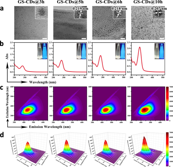
A sequence of GS-CDs at 170 °C utilizing GS as the one reactant at completely different response instances was synthesized, from 1 to 10 h, by way of regular hydrothermal synthesis. Excessive-resolution transmission electron microscopy (HRTEM) was used to characterize the scale and floor morphologies of GS-CDs@3 h, GS-CDs@5 h, GS-CDs@6 h, and GS-CDs@10 h at completely different hydrothermal response instances of three, 5, 6, and 10 h, respectively. All the GS-CDs confirmed good dispersity with even sizes, the place the diameter of GS-CDs@3 h was concentrated at 2.95 ± 0.72 nm, the diameter of GS-CDs@5 h was concentrated at 2.75 ± 0.66 nm, the diameter of GS-CDs@6 h was concentrated at 3.00 ± 0.64 nm, and the diameter of GS-CDs@10 h was concentrated at 3.24 ± 0.62 nm (Fig. 1a). The HRTEM photos proven within the higher proper nook of Fig. 1a introduced the crystal lattices of the 4 ready GS-CDs. GS-CDs@3 h had a mere crystal lattice, presumably because of the quick response time and incomplete nanostructure. GS-CDs@5 h had a sure lattice construction with a lattice spacing of 0.219 nm. GS-CDs@6 h and GS-CDs@10 h confirmed apparent lattices, with lattice spacings of 0.218 and 0.207 nm, respectively. This implied that with extended response time, the self-assembly of the GS molecules turned extra common, and the constructions of the GS-CDs turned extra full. In UV absorption spectra, the GS aqueous resolution had weak UV absorption, whereas the GS-CDs confirmed robust absorption at a wavelength of 280 nm (Fig. 1b and Extra file 1: Determine S1-1). The longer the response time, the upper the UV absorption peak. Mixed with IR spectroscopy evaluation, the absorption peak at 280 nm was primarily derived from the vibration and rotation of C = O (Extra file 1: Determine S1-2). The inset (Fig. 1b) is the photographs of the 4 ready GS-CDs below daylight (left) and UV gentle at 365 nm (proper), respectively. The GS-CDs with completely different response instances had robust blue-green fluorescence, and the brightness elevated barely with elevated response time.
Within the contour map of the three-dimensional fluorescence spectra of the 4 ready GS-CDs with completely different response instances, there was just one substance with fluorescent emission in every system, indicating a single product (Fig. 1c). As proven within the isometric projection of the three-dimensional fluorescence spectra (Fig. 1d), all of the 4 GS-CDs had excitation dependence. Underneath the optimum excitation wavelength of ~ 360 nm, the optimum emission wavelength was ~ 440 nm for the 4 GS-CDs. The longer the response time, the upper the obtained fluorescence depth of the GS-CDs. Within the fluorescence attenuation curves of the 4 GS-CDs (Extra file 1: Determine S1-3), three fluorescence lifetimes (τ) had been obtained, after fitted in keeping with a third-order decay exponential perform. It indicated that the CDs had three fluorescence facilities. The quantum yields of GS-CDs@3 h, GS-CDs@5 h, GS-CDs@6 h, and GS-CDs@10 h had been ~ 0.19%, ~ 0.14%, ~ 0.19%, and ~ 0.22%, respectively. X-ray photoelectron spectroscopy (XPS) was used to additional characterize and analyze the fundamental compositions and purposeful teams of the 4 GS-CDs. Within the XPS spectra, all of the 4 GS-CDs had two essential peaks at 285.1 and 531.6 eV, akin to the C 1s and O 1s parts, respectively (Extra file 1: Determine S1-4). The C 1s band had three peaks close to 288.5, 285.6, and 284.5 eV, which was attributed to the carbon peaks associated to C = O, C-O, and C-C. The O 1s band had two peaks close to 534.3 and 532.6 eV, which had been attributed to the oxygen peaks of C = O and C-O.
Characterization of GS-CDs below completely different synthesis circumstances. a TEM photos of the ready GS-CDs@3 h, GS-CDs@5 h, GS-CDs@6 h, and GS-CDs@10 h samples, respectively, the place the HRTEM photos inserted within the higher proper nook present the crystal lattices of the 4 GS-CDs. The dimensions bars had been 20 nm. b UV absorption spectra of the ready GS-CDs@3 h, GS-CDs@5 h, GS-CDs@6 h, and GS-CDs@10 h samples, the place the insets present the photographs of the 4 GS-CDs below daylight (left) and UV gentle at 365 nm (proper). c Contour maps exhibiting the three-dimensional fluorescence spectra of GS-CDs@3 h, GS-CDs@5 h, GS-CDs@6 h, and GS-CDs@10 h, respectively. d Isometric projections of the three-dimensional fluorescence spectra of GS-CDs@3 h, GS-CDs@5 h, GS-CDs@6 h, and GS-CDs@10 h.
Research have proven that the content material in typical conjugated fluorophore teams, corresponding to carbon-carbon double bonds, carbon-carbon triple bonds, or carbon-based π electrons in GS molecules may be very low [29]. The principle sources of UV absorption and fluorescence emission of the ready GS-CDs system didn’t consist of those basic conjugated constructions. The looks and enhance in UV and fluorescence could possibly be attributed to the cross-linking enhanced emission (CEE) impact [38]. Underneath a hydrothermal situation of 170 °C, and with speedy collisions between the particles, the free GS had been cross-linked via a buckling response, forming a carbon nucleus. Throughout the response, the principle tetracyclic triterpene construction of GS molecule confirmed few modifications, in keeping with the mass spectra evaluation of GS, GS-CDs@3 h, GS-CDs@5 h, GS-CDs@6 h, and GS-CDs@10 h (Extra file 1: Determine S1-5 and Desk S1-1). The energetic website beside the principle construction reacted with H2O, and the variety of C = O teams might enhance within the system (Extra file 1: Determine S1-2). With extended response time (3, 5, 6, and 10 h), the buckle response, in addition to the diploma of carbonization elevated. The constructions of the CDs turned denser and extra orderly (Fig. 1a), and extra C = O and C-O teams within the system turned mounted. Their vibrations and rotations had been restricted, leading to elevated radiation transition. Thus, the UV absorption and fluorescence emissions of the GS-CDs generated by non-classical conjugation had been enhanced [38, 39].
Inhibitory impact of GS-CDs on cells
The above outcomes demonstrated that below hydrothermal circumstances, and inside a sure response time, the GS molecules might self-assemble to type CDs. When response temperature (170 °C) was far beneath the edge temperature for the ring-opening response, the principle tetracyclic triterpene construction of GS couldn’t be destroyed in the course of the self-assembly course of. Thus, we proposed the self-assembled GS-CDs might retain biomedical exercise of GS to a sure extent. On this foundation, cell counting kit-8 (CCK-8) methodology was used to estimate cell viabilities of human neuroblastoma cells (SH-SY5Y), human cervical most cancers (HeLa), mouse microglial (BV2), rat adrenal pheochromocytoma (PC12), and human hepatoma (HepG2) cells. They had been incubated with completely different GS-CDs (GS-CDs@1 h, GS-CDs@2 h, GS-CDs@3 h, GS-CDs@4 h, GS-CDs@5 h, GS-CDs@6 h, GS-CDs@7 h, GS-CDs@8 h, GS-CDs@9 h, GS-CDs@10 h) for 48 h. The cell viabilities of HeLa, BV2, PC12, and HepG2 cells didn’t change considerably after incubation with GS-CDs below the identical circumstances (Extra file 1: Determine S2-1, S2-2, S2-3, and S2-4).
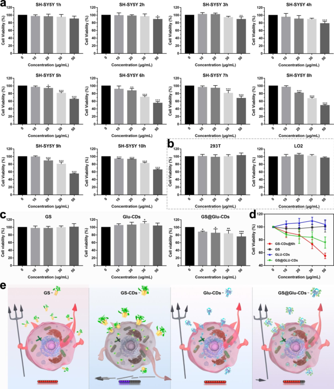
Observe that GS-CDs had few inhibitory results on human neuroblastoma cells (SH-SY5Y), when response time was 1–4 h (Fig. 2a). At a GS-CDs dosage of fifty µg·mL− 1, some cytostatic results appeared. When response time elevated to five h, GS-CDs began to show apparent inhibitory impact on SH-SY5Y cells. At a response time of 6 h, GS-CDs had a stronger inhibitory impact on SH-SY5Y cells. The cell viability charges had been ~ 87.56%, ~ 73.26% and ~ 55.05% at GS-CDs dosing concentrations of 20, 30 and 50 µg·mL− 1, respectively. The upper the dose, the decrease the cell viability. This prompt that the poisonous results of GS-CDs on SH-SY5Y cells had been concentration-dependent. When response time prolonged to 7–10 h, the inhibitory results of GS-CDs on SH-SY5Y cells had been just like GS-CDs@6 h. The 2 different repeated experiments had been principally an identical (Extra file 1: Determine S2-5). The experimental outcomes confirmed the ready GS-CDs had a really efficient inhibitory impact on SH-SY5Y cells. A low focus of GS-CDs (50 µg·mL− 1), their inhibition fee of SH-SY5Y cells achieved ~ 45.00%. Since response temperature (170 °C) was far beneath the edge temperature of the ring-opening response, the principle tetracyclic triterpenoid construction of GS couldn’t be destroyed. Subsequently, we proposed that GS-CDs retained the pharmaceutical exercise of the carbon-sourced GS, and launched largest variety of energetic substances into SH-SY5Y cells via endocytosis, with finest efficacy. As well as, because the substance of GS-CDs was pure plant essence, they confirmed good non-cytotoxicity to peculiar cells, corresponding to human renal epithelial (293T) cells and human regular liver (LO2) cells (Fig. 2b).
Contemplating the clearly increased inhibitory of GS-CDs on SH-SY5Y than different cells (HeLa, BV2, PC12, and HepG2 cells), there have been two speculations: (i) There are variations of energetic receptor in membrane floor amongst completely different tumor cells. GS-CDs might bind to sure particular receptors on the floor of SH-SY5Y cells, activating the tumor apoptosis pathway and thus exerting inhibitory results. (ii) Gene degree results: The incidence of tumors is the results of a number of genes, a number of steps, and a number of mutations. Mutations in numerous genes with completely different intensities result in the formation of various tumors. The addition of GS-CDs might change the construction of genetic DNA of SH-SY5Y cells, reverse the genetic traits of SH-SY5Y cells, and inhibit the tumor successfully.
Totally different inhibitory impact on SH-SY5Y cells
Moreover, below the identical circumstances we in contrast the inhibition effectivity of GS molecules and CDs with completely different construction/composition on SH-SY5Y cells, by way of in vitro cytotoxicity experiments (Fig. 2c and d, and 2e). Throughout the identical focus vary, GS had no inhibitory results on SH-SY5Y cells. It was inferred that GS in molecular state couldn’t be successfully taken up by way of cell transport and offered efficient drug impact. On opposite, because of the nano-size and nanostructure, GS-CDs could possibly be taken up by cells in giant numbers via endocytosis. Thus, GS-CDs confirmed a a lot increased inhibitory effectivity on SH-SY5Y cells than GS molecules. Second, molecule glucose of little medicinal exercise was used to organize widespread glucose carbon nanodots (Glu-CDs) by way of an identical hydrothermal response [14, 34, 40, 41]. When focus of Glu-CDs was 50 µg·mL− 1, viability of SH-SY5Y cells was weakly elevated. It indicated widespread Glu-CDs synthesized by non-drug molecules had little drug exercise. Lastly, GS was loaded onto the service Glu-CDs via supramolecular forces. Then drug molecule GS composite carbon nanodots (GS@Glu-CDs) had been obtained (Extra file 1: Fig. S2-6). On the identical focus of fifty µg·mL− 1, inhibitory impact (~ 75.69%) of GS@Glu-CDs on SH-SY5Y cells turned clearly however a lot lower than that of GS-CDs. It may be defined that after loaded on Glu-CDs, drug molecule GS might enter cell inside via endocytosis and had a dangerous impact on SH-SY5Y cells. Nevertheless, due to the nano-size of Glu-CDs, the loaded amount of bioactive GS was restricted, in addition to their inhibitory impact on SH-SY5Y cells. Thus, the development of nanodrug CDs composed of drug exercise molecules might open up new analysis concepts and utility instructions for nanomedicine.
Inhibitory impact of GS-CDs ready from 1–10 h and completely different CDs on SH-SY5Y cells. a In vitro cytotoxicity profiles of GS-CDs@1 h, GS-CDs@2 h, GS-CDs@3 h, GS-CDs@4 h, GS-CDs@5 h, GS-CDs@6 h, GS-CDs@7 h, GS-CDs@8 h, GS-CDs@9 h, and GS-CDs@10 h on SH-SY5Y cells. b In vitro cytotoxicity profiles of GS-CDs@6 h on 293T and LO2 cells. c In vitro cytotoxicity profiles of GS, Glu-CDs, and GS@Glu-CDs on SH-SY5Y cells. d Statistical chart summarizing the in vitro cytotoxicity profiles of GS-CDs@6 h, GS, Glu-CDs, and GS@Glu-CDs on SH-SY5Y cells. e Schematic photos exhibiting the cytotoxicity profiles of GS, GS-CDs, Glu-CDs, and GS@Glu-CDs on SH-SY5Y cells. Information had been imply ± s.d. (n = 6). *p < 0.05, **p < 0.01 and ***p < 0.001 relative to 0 µg·mL− 1, as analyzed by one-way evaluation of variance (ANOVA).
Intracellular distribution of GS-CDs in SH-SY5Y cells with time
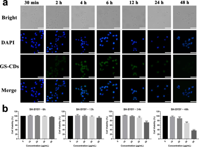
As a result of GS-CDs ready for six–10 h confirmed related inhibitory results on SH-SY5Y cells, GS-CDs@6 h had been chosen because the consultant nano-drug, and had been denoted as GS-CDs within the follow-up experiments. To additional discover the inhibitory impact of GS-CDs on SH-SY5Y cells, entry of GS-CDs into cells at completely different incubation instances was monitored by a fluorescent inverted microscope (Fig. 3a). Accompanied by its efficient medicinal results, the fluorescent GS-CDs system didn’t have to introduce different fluorescent substances for monitoring and labeling. With extended incubation time, SH-SY5Y cells step by step shrank and have become spherical. The SH-SY5Y cells step by step turned apoptotic with elevated time by the inhibition of GS-CDs. Within the fluorescence discipline, GS-CDs entered cells and had been primarily concentrated within the cytoplasm. Between 30 min and 6 h, fluorescence depth of the system step by step elevated, indicating the internalized variety of GS-CDs elevated with time. Between 6 and 12 h, fluorescence depth of the system decreased. This was presumably because of the reactions and construction destruction of GS-CDs throughout the cells. Fluorescence depth of the system was very weak after incubating for twenty-four h. By 48 h, little fluorescence was detected within the system. Whereas GS-CDs performed a medicinal function within the cells, they may be metabolized out via exocytosis. Fluorescence depth modifications within the incubation system indicated that inhibitory impact of GS-CDs on SH-SY5Y cells was time-dependent.
Cytotoxicity of GS-CDs in SH-SY5Y cells with time
The precise viability modifications of SH-SY5Y cell after incubating with GS-CDs for various instances had been investigated (Fig. 3b). When incubated for six and 12 h, the GS-CDs had a minimal inhibitory impact on the SH-SY5Y cells. When incubated for twenty-four h, at increased GS-CDs concentrations (50 µg·mL− 1), the inhibitory impact appeared. After 48 h of incubation, GS-CDs confirmed a powerful inhibitory impact on the SH-SY5Y cells, which was primarily per the outcomes above (Fig. 2a).
Fluorescent intracellular distribution, and cytotoxicity of GS-CDs in SH-SY5Y cells with time. a Fluorescence microscope photos of the SH-SY5Y cells and GS-CDs (100 µg·mL− 1) incubated at completely different instances (30 min, and a pair of, 4, 6, 12, 24, and 48 h). The dimensions bars had been 50 μm. b In vitro cytotoxicity profiles of the SH-SY5Y cells after incubating with the GS-CDs at completely different instances (6, 12, 24, and 48 h). Information had been imply ± s.d. (n = 6). *p < 0.05, **p < 0.01 and ***p < 0.001 relative to 0 µg·mL− 1, as analyzed by one-way ANOVA.
Bio-TEM evaluation was performed to check the mobile uptake and intracellular distribution of GS-CDs in SH-SY5Y, 293T and LO2 cells, respectively (Extra file 1: Determine S3-1). Many GS-CDs entered all of the cells and primarily existed in cytoplasm from 6 to 24 h. At 24 h, GS-CDs had been nonetheless ample in SH-SY5Y cells. In that case, some cells shrank, some cell membrane was damaged, and a few cell form was destroyed. It proved that GS-CDs inhibited SH-SY5Y cells successfully. Inside 6-12 h, GS-CDs additionally entered into 293T and LO2 cells, however little GS-CDs could possibly be discovered at 24 h. It implied GS-CDs had been principally metabolized inside 24 h in 293T and LO2 cells. What was extra, 293T and LO2 cells had regular morphology and good progress standing at 24 h. It prompt that GS-CDs had little poisonous impact on regular cells. Through the bio-TEM evaluation, it may be inferred GS-CDs with hydrophilic floor purposeful teams had been in a position to enter all these cells. GS-CDs possessed a excessive particular inhibitory impact on SH-SY5Y cells and little inhibitory impact on different cells.
Apoptosis and cell cycle of SH-SY5Y cells induced by GS-CDs
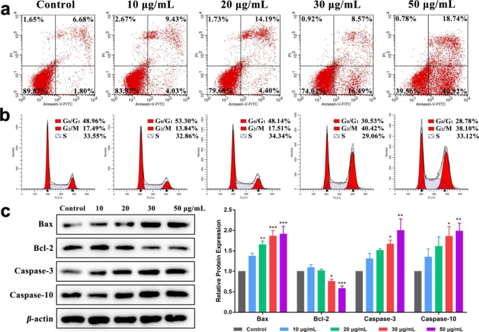
Move cytometry was used to detect apoptosis and cell cycle of SH-SY5Y cells after incubation at completely different GS-CDs concentrations after 48 h (Fig. 4a). When GS-CDs concentrations was 20, 30, and 50 µg·mL− 1, apoptosis fee of SH-SY5Y cells was ~ 18.59%, ~ 25.06%, and ~ 59.66%, respectively. The upper the administration focus, the extra important the apoptosis fee. It demonstrated when GS-CDs had been administered at excessive concentrations (for instance, 50 µg·mL− 1), and at a ample incubation time (48 h), an inhibition impact on SH-SY5Y cells could possibly be exerted.
When concentrations of GS-CDs had been 10 and 20 µg·mL− 1, the cell cycle of SH-SY5Y cells modified a bit (Fig. 4b). When focus of GS-CDs was elevated to 30 and 50 µg·mL− 1, SH-SY5Y cells in G0/G1 part decreased to ~ 30.53% and ~ 28.78%, respectively. Whereas in G2/M part, SH-SY5Y cells elevated to ~ 40.42% and ~ 38.10%, respectively. It indicated that GS-CDs might induce G2/M part arrest of SH-SY5Y cell cycle.
Apoptosis, cell cycle and expression of apoptosis-related proteins of SH-SY5Y cells induced by GS-CDs. a Apoptosis and b cell cycle charts of SH-SY5Y cells after incubating with completely different concentrations of GS-CDs (10, 20, 30, and 50 µg·mL− 1) for 48 h. c Results of corresponding completely different concentrations of GS-CDs on the expression of apoptosis-related proteins in SH-SY5Y cells
Impact of GS-CDs on the expression of apoptosis-related proteins in SH-SY5Y cells
The above apoptosis and cell cycle of SH-SY5Y cells confirmed that GS-CDs might inhibit tumor progress by inducing tumor cell apoptosis. Thus, we selected to detect the expression of apoptosis-related proteins in SH-SY5Y cells (Fig. 4c). Bax and Bcl-2 are homologous water-soluble associated proteins, which promote apoptosis in cells. Bax can antagonize the protecting impact of Bcl-2. Analysis confirmed that when the ratio of Bcl-2/Bax decreased, it could enhance the permeability of mitochondrial membrane, activate caspase apoptosis pathway and induce cell apoptosis [42]. Amongst them, caspase-10 is the downstream sign molecule of mitochondria that mediates apoptosis, and caspase-3 is the executor of apoptosis. Cell loss of life is inevitable after caspase-3 is activated [43]. Western-blot outcomes confirmed that in contrast with the management group, when the drug focus was 30 and 50 µg·mL− 1, the protein contents of Bax, caspase-3 and caspase-10 elevated considerably (P < 0.05, P < 0.01, P < 0.001). The content material of Bcl-2 protein decreased considerably (P < 0.05, P < 0.001). The above outcomes prompt that apoptosis of SH-SY5Y cells induced by GS-CDs could also be via inhibiting the up-regulation of Bcl-2, activating the caspase apoptosis pathway, activating and up-regulating the expression of apoptosis-related proteins caspase-3 and caspase-10. Lastly, it brought about SH-SY5Y cell loss of life.
Excessive inhibitory impact of GS-CDs on neuroblastoma and toxicity analysis in vivo
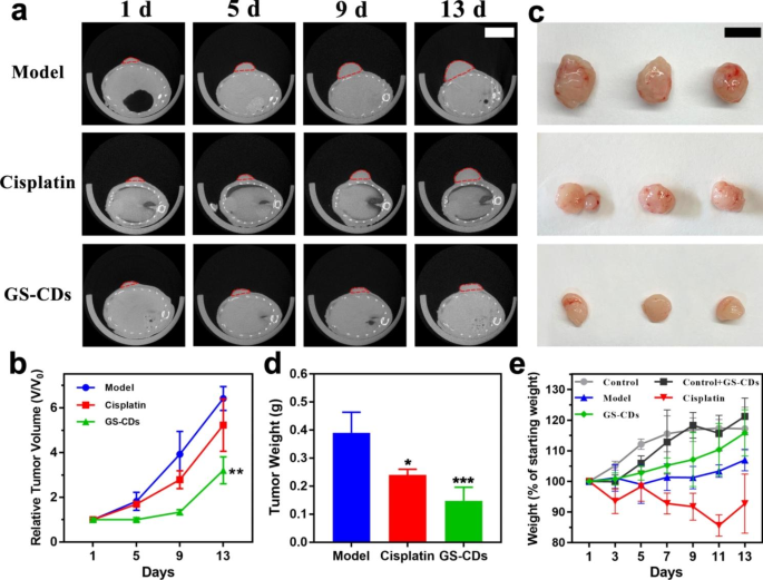
To guage the inhibited skill of GS-CDs on human neuroblastoma in vivo, we employed BALB/c nude mice bearing human neuroblastoma tumors because the animal mannequin. The antineoplastic cisplatin for the scientific therapy of neuroblastoma was administered to tumor-bearing mice because the constructive group. The true-time photographs of mice progress within the mannequin, cisplatin, and GS-CDs group had been recorded fastidiously (Extra file 1: Determine S5-1, S5-2, and S5-3). Utilizing a small animal CT imaging system, CT scans had been carried out to look at the expansion of the mice tumors on the first, fifth, ninth, and thirteenth days throughout one experiment cycle (Fig. 5a). The statistic relative tumor quantity of the mannequin, cisplatin, and GS-CDs group, was calculated by the CT scans of the most important tumor sections (Fig. 5b). With time, the tumor progress pattern of mannequin group was apparent, and tumor progress in cisplatin group was slower than within the mannequin group. The tumor progress fee of mice in GS-CDs group was the slowest and the tumor quantity was the smallest. On the finish of thirteenth day of experimental interval, mice in every group had been dissected and tumor comparisons had been made (Fig. 5c). The tumor quantity and statistic tumor weights of the GS-CDs group had been considerably smaller than the opposite two teams (Fig. 5d). The above knowledge confirmed that the ready GS-CDs might successfully inhibit the expansion of human neuroblastoma and produce substantial healing results in vivo.
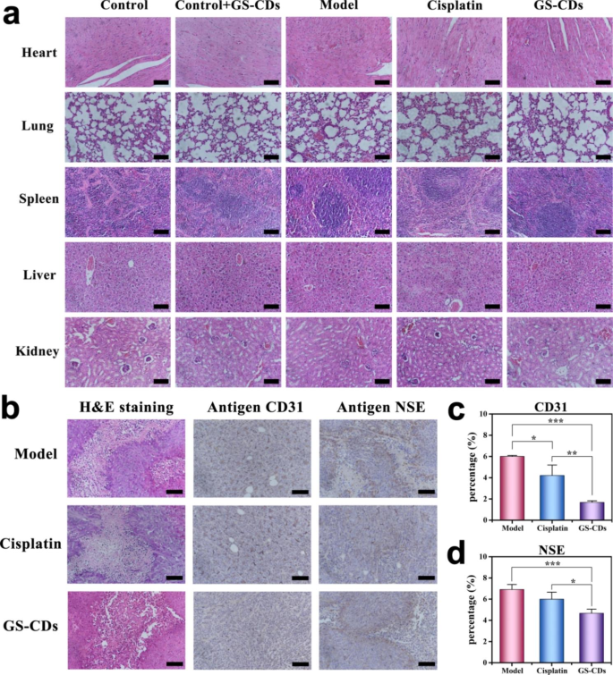
In the meantime, statistical diagrams for weight modifications of mice within the management, management + GS-CDs, mannequin, cisplatin, and GS-CDs group had been additionally recorded to evaluate the toxicity of GS-CDs (Fig. 5e). In comparison with the burden of mice in management group, weight of mice in management + GS-CDs group was barely decrease on the preliminary stage. With extra time, weight of mice in management + GS-CDs group exceeded that of management group. This proved that the ready GS-CDs had virtually no poisonous results on organism. In comparison with the burden of mice in mannequin group, weight of mice in GS-CDs group was very important. This additionally confirmed that GS-CDs didn’t produce poisonous results throughout neuroblastoma therapy. Attractively, as a result of it might successfully inhibit tumor progress and enhance well being of mice, the burden of mice in GS-CDs group was near the management group on the thirteenth day. The load lack of mice within the cisplatin group was probably the most. The H&E staining of the principle organs of mice in every group outcomes confirmed that few apparent organ abnormalities had been noticed within the pathological photos of mice in cisplatin group (Fig. 6a). Experimental observations discovered that the mice injected with cisplatin had been in a depressed state in comparison with mice in different teams. They had been reluctant to eat for a time frame. This was presumably because of the extreme irritation of cisplatin on the intestines and stomachs of the mice, leading to important weight reduction. The above outcomes indicated that the GS-CDs had excessive biocompatibility within the therapy course of and could possibly be used safely and successfully to deal with neuroblastoma. It must be emphasised that human neuroblastoma is principally dangerous to infants and younger youngsters with low immunity. For these youngsters who must be fastidiously protected throughout therapy, the unwanted side effects attributable to the nano-drug GS-CDs primarily based on pure natural extracts could be enormously diminished. Thus, the acceptance charges for infants and younger youngsters could be a lot increased. It could be extra conducive to the therapy of the illness.
Inhibitory impact of GS-CDs on neuroblastoma in vivo. a Tumor progress of the mice tumors within the mannequin (high), cisplatin (center), and GS-CDs (backside) group on the first, fifth, ninth, and thirteenth days as noticed by CT scans (n = 3), the place the size bar was 1 cm. b Statistic charts exhibiting the relative tumor volumes of the mice within the mannequin, cisplatin and GS-CDs group, as calculated by the CT scans of the most important tumor sections. Information had been imply ± s.d. (n = 3). c Photographs of the dissected mice tumors within the mannequin (high), cisplatin (center) and GS-CDs (backside) group at thirteenth day (n = 3), the place the size bar was 1 cm. d Statistic chart exhibiting the tumor weight of the mice within the mannequin, cisplatinand GS-CDs group. e Statistical weight diagrams of the mice in management, management + GS-CDs, mannequin, cisplatin, and GS-CDs teams. Information had been imply ± s.d. (n = 3). *p < 0.05, **p < 0.01 and ***p < 0.001 relative to the mannequin group, as analyzed by one-way ANOVA.
To estimate pathological injury of GS-CDs on tumors, the dissected neuroblastoma of mice within the mannequin, cisplatin, GS-CDs group had been stained and analyzed (Fig. 6b-d). As a result of human neuroblastoma tumor cells had been malignant and grew rapidly, sizes of tumors within the management group had been very giant on the finish of the check interval. The offered blood and vitamins had been inadequate for the tumor, inflicting necrosis of the cells inside tumor. There was ample blood provide outdoors tumor, and the tumor might preserve speedy progress. As noticed within the H&E (hematoxylin-eosin staining) picture, tumor cells in every group had a sure diploma of harm, together with within the mannequin group. Neovascularization is a vital situation for tumor progress, and antigen CD31 (endothelial cell adhesion molecule) was employed to label the vascular endothelial cells in tumor. The expression of CD31 in GS-CDs group was clearly decrease than the opposite two teams. As well as, antigen neuron-specific enolase (NSE) is a marker of neuroblastoma. In comparison with the opposite teams, expression degree of NSE in GS-CDs group was the bottom.
It was worthy to notice that since we didn’t modify the surfaces of GS-CDs with sensible industrial fluorescent agent or distinction agent, it was arduous to review the precise biodistribution of GS-CDs in animals. We’ve got checked the literatures inside final ten years to invest the fates of GS-CDs in animals (Extra file 1: Desk S6-1) [16, 44,45,46,47,48].
Stained sections of essential organs and neuroblastoma tissues of mice. a H&E staining diagrams of the principle organs (coronary heart, lungs, spleen, liver, and kidneys) of mice within the management, management + GS-CDs, mannequin, cisplatin, and GS-CDs teams, the place the size bars had been 100 μm. b H&E (left), antigen CD31 (center), and antigen NSE (proper) stained neuroblastoma tissues of mice at thirteenth day within the mannequin (high), cisplatin (center) and GS-CDs (backside) group, the place the size bars had been 100 μm. c Statistic comparability chart of the antigen CD31 immunohistochemical stained neuroblastoma tissues of the mice within the mannequin, cisplatin and GS-CDs group. d Statistic comparability chart of the antigen NSE immunohistochemical stained neuroblastoma tissues of the mice within the mannequin, cisplatin, and GS-CDsgroup. Information had been imply ± s.d. (n = 4). *p < 0.05, **p < 0.01 and ***p < 0.001 relative to the mannequin group or cisplatin group, as analyzed by one-way ANOVA.


