The preparation and characterization of SC@DS NPs
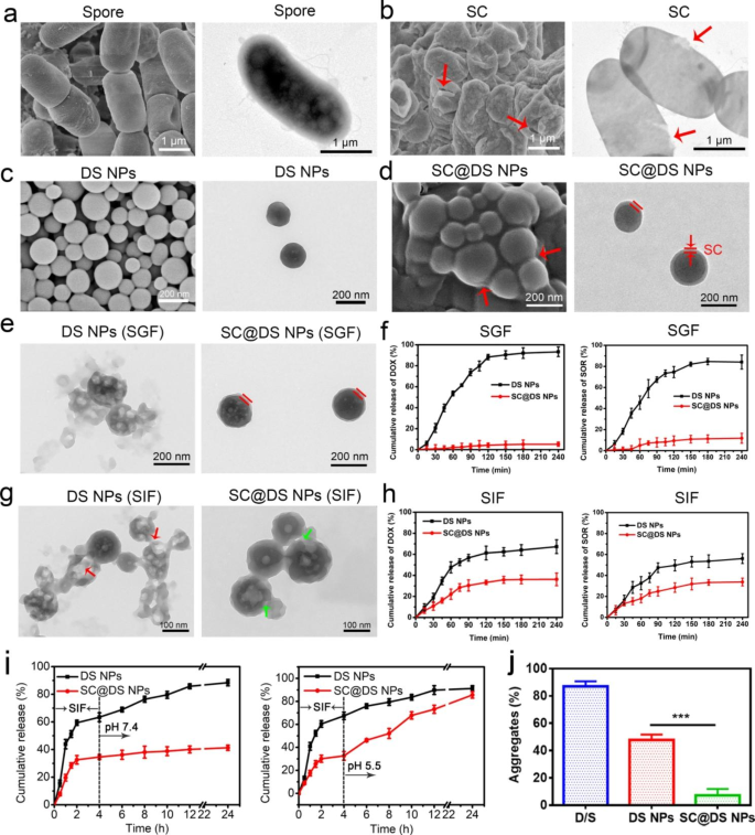
Firstly, the morphology of BC and spore was characterised by Scanning Electron Microscopy (SEM) and Transmission Electron Microscopy (TEM), respectively. As might be seen, BC with the particle measurement of three ~ 5 μm was in rod-shaped (Further file 1: Fig. S1), whereas spores of 1 ~ 2 μm with plump and integrity form have been separated efficiently from BC (Fig. 1a). Furthermore, Gram straining additionally confirmed that the profitable cultivation of BC and isolation of spores (Further file 1: Fig. S2). Subsequently, so as to put together spore capsid (SC), BC spores have been incubated with completely different focus sodium chloride (NaCl) resolution. In contrast with the graceful floor of spores that dispersed in saline (Fig. 1a), slight fold was noticed on the floor of spores after being incubated for two h in 5% NaCl (Further file 1: Fig. S3a), and the shrinkage diploma of spores elevated with the NaCl focus growing (Further file 1: Fig. S3b). Due to this fact, on this examine, spores have been dispersed in 20% NaCl to acquire the pure SC. As proven in Fig. 1b, extra extreme morphology shrinkage of spores was noticed. Furthermore, the TEM picture confirmed the clear breaches within the spores the place the crimson arrows level, which might trigger the primary content material was launched from spores. This phenomenon indicated that SC was remoted efficiently from the spores. Given the excessive resistance of SC to the tough atmosphere [17], we speculated that SC might be served as a car for delivering the nanoparticles (NPs) by oral administration. After which SC was damaged by Ultrasonic Homogenizer to arrange SC particles. As proven within the Further file 1: Fig. S4, BC introduced lamellar construction, which was conducive to the preparation and formation of NPs and medicines loading.
Preparation and characterization. SEM and TEM photos of (a) spores and (b) SC, respectively. The SEM and TEM photos of (c) DS NPs and (d) SC@DS NPs, respectively. (e) The morphologies of DS NPs and SC@DS NPs have been investigated after 2 h incubation in SGF. (f) The cumulative DOX and SOR launch of DS NPs and SC@DS NPs in SGF. (g) The morphology adjustments of DS NPs and SC@DS NPs after 4 h incubation in SIF. (h) The cumulative launch quantity of SOR and DOX in DS NPs and SC@DS NPs teams after being incubated in SIF for 4 h. (i) The discharge habits of medication from DS NPs and SC@DS NPs after being incubated in a buffer with step by step various pH for over 24 h. (j) The proportion of particles-mucin aggregation fashioned in simulated mucus resolution for various preparations at 37℃
Moreover, the technique of mixture of two medicine can be utilized to reinforce the therapeutic impact [31,32,33]. Accordingly, as a proof of idea, the water-soluble DOX and hydrophobic SOR have been chosen to type DS NPs by the nanoprecipitation methodology [30]. Surprisingly, as proven in Fig. 1c and Further file 1: Fig. S5, the DS NPs have been of fine morphology and nicely dispersion. The UV absorption peak at 266 nm (crimson arrow) and 480 nm (black arrow) of the DS NPs was in line with the utmost absorption peak of free SOR and DOX, respectively, which additional proved that the composition of DS NPs was DOX and SOR (Further file 1: Fig. S6). Subsequently, as proven in Fig. 1d and Further file 1: Desk S1, in contrast with DS NPs, the thick coating was noticed on the floor of SC@DS NPs, which confirmed that SC@DS NPs have been synthesized efficiently after being extruded for 13 instances. Furthermore, the potent wrapping of SC onto the floor of DS NPs was additional demonstrated by the slight particle measurement growing and floor cost variation in contrast with the uncoated DS NPs (Further file 1: Fig. S7 and S8). Notably, the protein composition of SC@DS NPs was much like that of SC, indicating that SC coating course of might hardly have an effect on the floor composition of SC (Further file 1: Fig. S9). With extension of the storage time at room temperature, the negligible adjustments of particle measurement and zeta potential illustrated the superior stability of SC@DS NPs (Further file 1: Fig. S10 and S11). Moreover, the soundness of SC@DS NPs in PBS, serum and cell tradition media additionally was evaluated. As proven in Further file 1: Fig. S12, SC@DS NPs have been in an excellent dispersion in varied organic media, indicating their good biocompatibility and organic stability.
Contemplating that spore coat is the important thing issue of spore resistance to the tough acidic atmosphere, we speculated that SC was comparatively steady in abdomen. So as to examine whether or not SC might defend DS NPs from the intense abdomen situation, the DS NPs and SC@DS NPs have been incubated within the simulated gastric fluid (SGF), respectively. After 2 h incubation, their morphologies have been noticed by TEM photos. As proven in Fig. 1e, DS NPs have been destroyed whereas SC@DS NPs might keep the whole form, which urged that SC might be served because the protecting car. As well as, through the incubation in SGF, the drug launch of DOX and SOR was additionally evaluated. As might be seen in Fig. 1f, the discharge quantities of DOX and SOR in DS NPs have been near 100%. Quite the opposite, the negligible launch quantity was detected in SC@DS NPs, which additional testified the protecting perform of SC. Moreover, the drug launch properties and the morphology adjustments of DS NPs and SC@DS NPs have been evaluated after each of the NPs have been incubated in stimulated intestinal fluids (SIF) for 4 h, respectively. As proven in Fig. 1g, most DS NPs have been damaged (crimson arrow) and couldn’t keep an entire morphology. Though the SC@DS NPs might maintain a good condition, there have been nonetheless some slight damages due to the presence of intestinal particular enzymes, which might result in a low drug launch price of about 30% within the SIF (Fig. 1h). After which, after the DS NPs and SC@DS NPs have been pre-incubated in SIF for 4 h, they have been transferred to the answer of pH 7.4 and pH 5.5 for one more incubation time. As proven in Fig. 1i, the excessive launch price of DS NPs was detected in each pH 7.4 and pH 5.5. Nevertheless, for the SC@DS NPs, there was virtually no drug launch after being incubated in pH 7.4 for 20 h, whereas the sustained drug launch was noticed after they have been transferred to the pH 5.5 resolution. This end result urged that SC@DS NPs might obtain a sustained drug launch within the tumor microenvironment.
Notably, mucus barrier might lower the effectivity of the intestinal epithelial absorption of NPs [34]. We additional analyzed the precise protein parts of SC by LCMSMS (nanoLC-QE). As proven in Further file 1: Desk S2, the SC incorporates quantity of cysteine, which might result in the excessive mucus penetration of SC. As everyone knows, mucus consists of the advanced biochemical compositions, which might adsorb a variety of molecules and particles, together with medicine, and different probably dangerous entities [35, 36]. As proven in Fig. 1j, a excessive aggregates price was detected within the D/S remedy group, which was as a result of the small molecule medicine might be trapped and shortly eliminated by intestinal mucus. Amongst these teams, SC@DS NPs confirmed a low mixture price with a share of seven.2% ± 4.7%, which might be interpreted as that SC containing quantity of cysteine might cleave the disulfide bonds between mucus glycoproteins, growing the intestinal mucus penetration of DS NPs [37, 38]. Moreover, the vertical distribution of NPs (crimson) on cell monolayer have been noticed by Confocal Laser Scanning Microscopy (CLSM) utilizing Z-axial scanning (Further file 1: Fig. S13). The fluorescence alerts of DOX and DS NPs remedy teams exhibited a powerful correlation with the mucus layer whereas only a few alerts have been overlaid with the mucus for SC@DS NPs, indicating that a big proportion of SC@DS NPs might escape the trapping of mucus. Moreover, we subsequent investigated whether or not the variations are attributed to alterations in lure or permeability of NPs on the mucosal floor. The numerous separation of crimson and inexperienced fluorescence was noticed within the SC@DS NPs, indicating its superior capability to flee from mucus trapping and good permeability (Further file 1: Fig. S14).
Uptake mechanism and transepithelial transport of SC@DS NPs
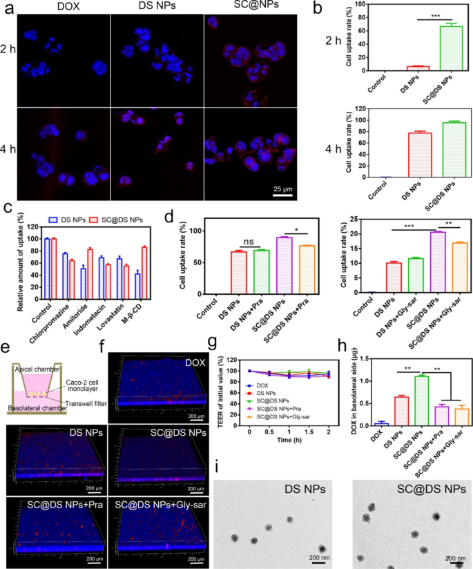
Environment friendly epithelium uptake and transepithelial transport play a necessary position to the oral NPs supply [39]. Firstly, the cytotoxicity of SC and SC@DS NPs was evaluated on Caco-2 cells, respectively. When the concentrations of SC and SC@DS NPs reached 500 µg/mL, the cell viability price was about 95%, indicating that the drug service system has good biocompatibility and no apparent toxicity on Caco-2 cells (Further file 1: Fig. S15). Notably, we discovered that the cell uptake price on Caco-2 cells was considerably improved by SC@NPs in contrast with the opposite handled teams (Fig. 2a, b and Further file 1: Fig. S16). This phenomenon is as a result of SC incorporates some goal proteins which might bind to the receptors in epithelium. Intrigued by this promising end result, we additional investigated the endocytosis mechanism of SC@DS NPs. As proven in Fig. 2c, amiloride and M-β-CD have been served because the non-specific inhibitors for macropinocytosis and pathway inhibitor of disruptor of lipid raft, respectively, which might considerably cut back the mobile uptake of the DS NPs handled group. Furthermore, chlorpromazine and lovastatin have been served because the inhibitors of clathrin-mediated endocytosis, and caveolae-mediated endocytosis pathways, respectively. Apparently, SC@DS NPs displayed a marked inhibition impact of mobile uptake after being handled with chlorpromazine (11% discount) and lovastatin (11.9% discount), respectively. These outcomes indicated that the mobile uptaken effectivity of SC@DS NPs was concerned within the a number of mobile pathways similar to lipid raft and caveolae-mediated uptake, clathrin-dependent endocytosis in addition to macropinocytosis. As anticipated, this non-specificity of endocytosis pathway might contribute to the advance of their mobile uptake effectivity.
Research of uptake mechanism and transepithelial transport. (a) Cell uptake of free DOX, DS NPs and SC@DS NPs at 2 and 4 h incubation, respectively. (b) The results of movement cytometry statistical evaluation after the Caco-2 cells being incubated with DS NPs and SC@DS NPs after 2 and 4 h incubation, respectively. (c) The measurement of relative uptake on Caco-2 cells after being incubated with the completely different endocytosis inhibitors. (d) The influences of Pra and Gly-sar on the mobile uptake of DS NPs and SC@DS NPs, respectively. (e) Institution of Caco-2 cell monolayer fashions. (f) The CLSM photos of various NPs on Caco-2 cell monolayers from apical to basolateral aspect. (g) TEER values of Caco-2 cell monolayers earlier than and after completely different incubations. (h) The quantity of transepithelial transport of various teams within the basolateral chamber. (i) The TEM photos of DS NPs and SC@DS NPs within the basolateral chamber
Moreover, we additionally studied the precise receptor-mediated endocytosis that associated to the excessive uptake of SC@DS NPs. The beforehand research claimed that the monocarboxylate transporter-1 (MCT1) was the primary receptor within the transport of short-chain fatty acid (SCFA), similar to butyrate, lactate and propionate [40]. As proven in Fig. 2d, the mobile uptake of SC@DS NPs was evidently depressed by pravastatin (Pra) generally known as the inhibitor of MCT1. This end result positively supported that MCT1 performed a significant position for the endocytosis of SCFA-functionalized nanovehicles. In addition to, the elevated uptake price in SC@DS NPs group is perhaps related to the additional activation of the opposite pathways. As reported, SC incorporates quantity of tyrosine, which could enhanced the receptor-mediated endocytosis by the oligopeptide transporter (PePT1) [41]. The glycyl-sarcosine (Gly-Sar), because the inhibitor of the PePT1 receptor, might considerably lower the uptake of SC@DS NPs. With out particular interplay with MCT1, SC@DS NPs nonetheless confirmed the excessive mobile uptake price.
Subsequently, the in vitro transepithelial transport of various NPs was evaluated by the Caco-2 cell monolayers (Fig. 2e). To our shock, as proven in Fig. 2f, the fluorescence of SC@DS NPs remedy group was embedded within the cell monolayer penetrating to the basal aspect of the monolayer. Whereas the fluorescence of different teams was floating on the monolayer, and the penetrating phenomenon of those NPs was hardly noticed. Nevertheless, SC@DS NPs hardly achieved environment friendly transepithelial transport when the cell monolayers have been incubated with the completely different pathway inhibitors, respectively. This end result was interpreted by the truth that the transport effectivity of SC@DS NPs might be enhanced by the receptor mediated endocytosis. Moreover, the transepithelial electrical resistance (TEER) values of cell monolayer have been practically unchanged earlier than and after the completely different remedies (Fig. 2g). And the tight junction protein was introduced on the border of epithelial cells and the apical mobile membrane, and the optimistic expression of Occludin in sections is marked crimson colour (Further file 1: Fig. S17). Apparently, in line with the management group, the annular and steady crimson fluorescence was noticed across the nucleus within the SC@DS NPs group. Nevertheless, the discontinuous crimson fluorescence was detected within the different handled teams. These outcomes all proved that the permeability of SC@DS NPs was brought on by receptor-mediated transcellular transport moderately than breakage of the integrity of cell monolayers. The quantity of transcellular transport of SC@DS NPs was measured in basolateral chamber, exhibiting 2.6-fold and a couple of.9-fold (P < 0.05) larger than that of the group pre-treatment with Pra and Gly-sar, respectively (Fig. 2h). In the meantime, the morphology of various NPs in basolateral chamber was detected by TEM. As proven in Fig. 2i, the sting of DS NPs was tough whereas the floor of SC@DS NPs with integrity was easy, which indicated that SC@DS NPs had good stability after they have been transported throughout the epithelium.
Analysis of anti-tumor effectivity in vitro
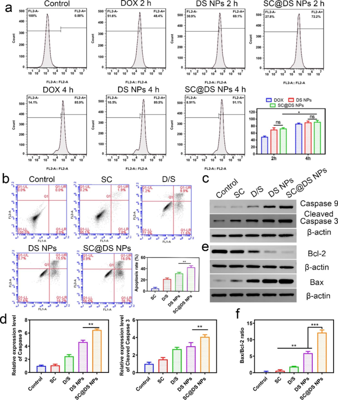
The in vitro anti-tumor effectivity was investigated by the human colon tumor cell line SW620. As proven in Fig. 3a and Further file 1: Fig. S18, there have been no vital variations between DS NPs and SC@DS NPs remedy teams after 2 and 4 h incubation, respectively. This end result urged that SC might solely enhance the intestinal epithelial transport effectivity because of the particular receptors, and it couldn’t carry out apparent mobile uptake in tumor cells. Nevertheless, the cell uptake of DS NPs and SC@DS NPs was observably larger than that of DOX with the incubation time. In addition to, the apoptosis stage of various group in SW620 cells was additionally detected. As proven in Fig. 3b, there have been extra apoptotic cells in SC@DS NPs group with an apoptotic price of 42.7 ± 3.0%, which was considerably larger than that of DS NPs remedy group (21.7 ± 2.2%) and D/S remedy group (21.5 ± 2.1%). This end result was attributed to the truth that SC additionally might induce a light apoptosis on tumor cells. The apoptosis stage was additional decided by Western blotting evaluation. As proven in Fig. 3c, d and Further file 1: Fig. S19, a big enchancment within the apoptosis proteins similar to caspase-9 and cleaved caspase-3 was noticed after incubation with SC@DS NPs, indicating that SC@DS NPs might induce extreme cell apoptosis. Furthermore, the extent of tumor cell apoptosis can also be regulated by the antiapoptotic protein bcl-2 and the apoptotic protein bax [42]. The expression of bcl-2 and bax and the semi-quantitative evaluation of bcl-2/bax ratio for every group have been proven in Fig. 3e, f and Further file 1: Fig. S20. For the SC@DS NPs remedy group, the bax/bcl-2 ratio was remarkably decrease than that of the opposite teams, indicating that this preparation might improve the method of cell apoptosis.
Mobile uptake and apoptosis evaluation in SW620 tumor cells. (a) Quantitative dedication of cell uptake quantities at no cost DOX, DS NPs and SC@DS NPs at 2 and 4 h incubation, respectively. (b) Apoptosis stage of SW620 cells after being handled with D/S, DS NPs, SC and SC@DS NPs, respectively. (c) The caspase-9 and cleaved-caspase-3 protein ranges of SW620 cells and (d) semi-quantitative evaluation of various teams. (e) The bax/bcl-2 ratio evaluation
Intestinal absorption in vivo
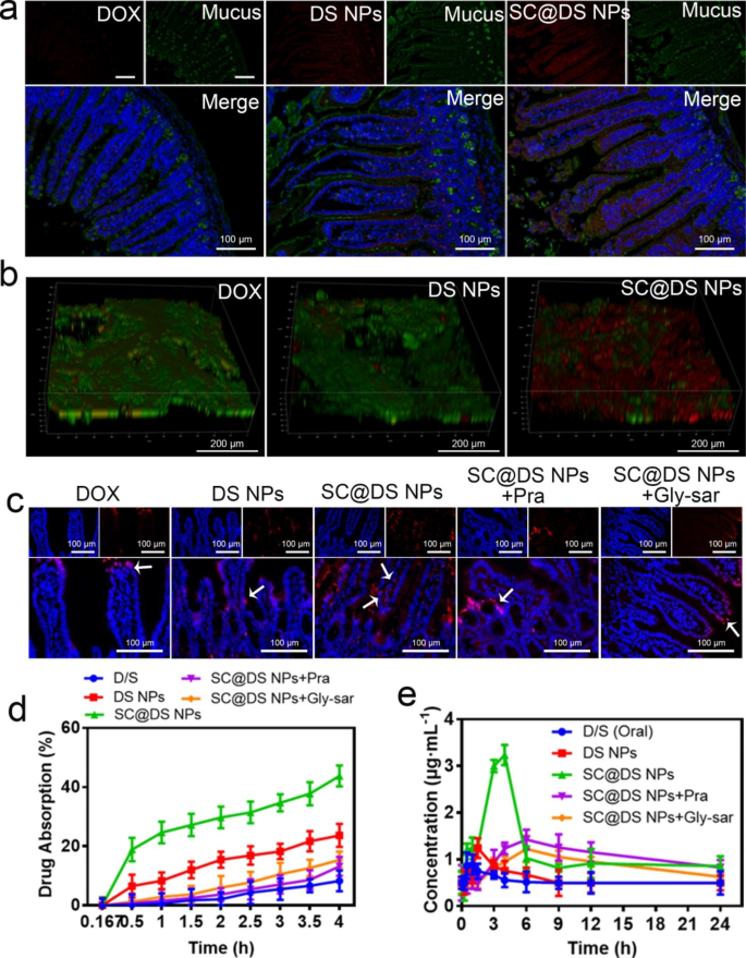
The earlier examine claimed that the sulfhydryl teams of cysteine on the floor of spores might cleave the disulfide bond between mucin glycoproteins and enhance the intestinal mucus penetration of NPs [16]. As proven in Fig. 4a and Further Fig. S21, low fluorescence depth was noticed within the DOX-treated group, which indicated that DOX couldn’t penetrate the mucus. For the DS NPs-treated group, although the marked fluorescence sign was detected, the intestinal epithelial mobile uptake exhibited a powerful correlation with the mucus layer, indicating {that a} bigger proportion of DS NPs was trapped throughout the mucus. This end result was additionally demonstrated within the in vitro particles-mucin aggregation experiment. Quite the opposite, the upper fluorescence depth was noticeably noticed within the intestinal villi, whereas only a few fluorescence alerts have been overlaid with the mucus. Furthermore, as proven in Fig. 4b, the 3D photos of the intestinal tissues with completely different remedy additionally proved that SC@DS NPs might dramatically keep away from mucus trapping and enhance the uptake effectivity of intestinal epithelial cells. This phenomenon might be interpreted because the protecting useful of the SC.
Analysis of intestinal absorption and pharmacokinetic parameters. (a) The mucus penetration impact of various NPs. (b) The everyday 3D CLSM photos of the completely different NPs distribution in mucus. (c) Consultant fluorescence photos of gut after the mice have been handled with free DOX, DS NPs, SC@DS NPs, SC@DS NPs + Pra and SC@DS NPs + Gly-sar by gavage, respectively. (d) Analysis of the intestinal absorption of various teams by the in situ intestinal circulation and absorption mannequin. (e) Drug focus in plasma after oral administration with completely different teams at a dose equal to 30 mg kg− 1 of DOX.
For additional investigating the in vivo intestinal absorption of various NPs, the mice have been orally handled with free DOX, DS NPs and SC@DS NPs for 4 h, respectively. As proven in Fig. 4c, the weak crimson fluorescence sign was detected within the intestinal villi after the mice have been handled with DS NPs, indicating the poor absorption of DS NPs. As anticipated, the sturdy fluorescence sign was clearly noticed at each the epithelial cells and basolateral aspect of the epithelium within the SC@DS NPs remedy group. Apparently, when the SC@DS NPs handled group was pre-incubated with Pra and Gly-sar, respectively, they exhibited a weak fluorescence sign within the epithelial cells. This end result demonstrated that SC@DS NPs have been effectively absorbed on the epithelium and efficiently transported into the lamina propria by the receptor-mediated endocytosis. Subsequently, the analysis of in situ intestinal absorption and circulation was carried out by the ex vivo ligated intestinal loop mannequin. As proven in Fig. 4d, the quantity of drug absorption of SC@DS NPs was 43.8% ± 3.5%, which was considerably larger than that of DS NPs with the drug absorption of 23.6% ± 3.9%. Nevertheless, when the transcellular pathway was inhibited by Pra or Gly-sar, the intestinal epithelial absorption of SC@DS NPs was 13.2% ± 3.1% and 15.4% ± 2.8%, respectively. These outcomes urged that SC@DS NPs might enhance the effectivity of transepithelial transport and absorption, which was the excellent prerequisite to reinforce the therapeutic impact.
Given the promising traits of SC@DS NPs, we additional evaluated the oral relative bioavailability after administration with D/S, DS NPs, SC@DS NPs, SC@DS NPs + Pra and SC@DS NPs + Gly-sar, respectively. The plasma focus time curves and pharmacokinetic parameters have been proven in Fig. 4e and Further file 1: Desk S3. The Cmax worth of DS NPs was 1.24 ± 0.35 µg mL− 1 at 1.5 h, which was decrease than that of the opposite remedy teams. This end result was primarily because of the poor stability of DS NPs after they handed by means of the tough abdomen situations. Notably, the SC@DS NPs handled group achieved the very best relative bioavailability (Frel), which was 5.6-fold larger than the values obtained by the DS NPs remedy. As well as, when the mice have been pre-treated with the receptor inhibitors and adopted by.
remedy with SC@DS NPs, their relative bioavailability was remarkably decreased. Taken collectively, SC@DS NPs with the superior mucus-penetrating and mobile uptake capabilities might be served as a priceless drug supply system, enhancing the medicine bioavailability.
In vivo distribution and anti-tumor efficacy
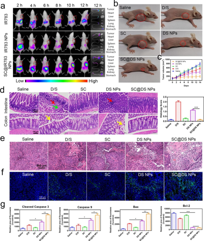
For investigating the in vivo distribution of NPs, a near-infrared dye IR783 was employed to exchange DOX to type the IR783 NPs self-assembly. As proven in Fig. 5a, the fluorescence sign notably gathered into the abdomen at 2 h for the free IR783 and IR783 NPs remedies, respectively. As time continued to increase, the fast lower of fluorescence distribution in vivo was clearly noticed. In contrast, after the mice have been handled with SC@IR783 NPs, the tumor website exhibited a powerful fluorescence sign at 8 h and remained apparent fluorescence till 12 h, whereas the opposite handled teams displayed virtually negligible fluorescence sign. This phenomenon of the NPs real-time distribution of the key organs and intestinal tissues might be interpreted as follows: (i) SC might defend NPs from the tough gastrointestinal tract atmosphere; (ii) SC might effectively improve the transepithelial transport capability of NPs by the a number of transport pathways, which might enhance the quantity of NPs that entered the bloodstream. These promising in vivo distribution traits of NPs would possibly result in a superior anti-tumor efficacy.
The in vivo distribution and remedy efficacy of various NPs. (a) In vivo imaging of SW620 tumor-bearing mice on the preset time factors. (b) Consultant photos of mice after completely different remedies. (c) Modifications in tumor quantity throughout remedy. (d) Consultant H&E photos of intestinal and colorectal tissues and histology rating after completely different remedies (Pink arrow: focal lymphocytic infiltration; Yellow arrow: desquamation of epithelial cells, Inexperienced arrow: seen connective tissue hyperplasia within the lamina propria); Scale bar = 100 μm. (e) H&E staining of tumor tissues from completely different remedies (Scale bar = 500 μm). (f) TUNEL staining of tumor tissues on the finish of every remedies (Scale bar = 100 μm). (g) Quantitative evaluation of apoptosis and anti-apoptosis associated proteins together with cleaved caspase-3, caspase-9, bax, and bcl-2 in numerous remedies (imply ± SD, n = 6, *P < 0.05, **P < 0.01, ***P < 0.001). The completely different formulations have been orally administered to rats at a dose equal to 30 mg kg− 1 of DOX and 5 mg kg− 1 of SOR, respectively. Important variations have been assessed utilizing two-way evaluation of variance (ANOVA) with a number of comparisons or utilizing the t-test for comparability of two teams
Subsequently, the in vivo tumor inhibition effectivity was investigated after remedy with saline, D/S, SC, DS NPs and SC@DS NPs for 14 days, respectively. As proven in Further file 1: Fig. S22, the physique weight acquire of D/S and SC handled teams was gradual as a result of DOX had cardiotoxicity, whereas SC had solely weak therapeutic impact. The physique weight of the mice handled with SC@DS NPs was elevated clearly, which urged the nice biocompatibility and remedy impact of SC@DS NPs. Furthermore, the tumor volumes of every mouse have been recorded to guage the remedy efficacy of various teams. As depicted in Fig. 5b, c and Further file 1: Fig. S23, no apparent tumor inhibition was noticed in saline and SC remedy teams. By evaluating with the tumor volumes of D/S, DS NPs and SC@DS NPs remedy teams, the anti-tumor effectivity of SC-coated NPs on mice was higher than that in free medicine and NPs group. As soon as the drug was ready to the NPs after which functionalized with SC, the tumor inhibition efficacy was improved. These outcomes confirmed that SC with good biosafety might be used because the automobiles for drug supply and had a superb protecting impact on NPs, which may enhance the soundness of NPs within the advanced GIT situations and enhance the anti-tumor impact.
Then the pathological options of small gut and colon tissues have been evaluated by the histology rating adjustments in line with the precise parameters (Further file 1: Desk S4). As proven in Fig. 5d, the hematoxylin and eosin (H & E) assay urged that D/S and DS NPs might induce the inflammatory response similar to focal lymphocytic infiltration; desquamation of epithelial cells and visual connective tissue hyperplasia within the lamina propri. Nevertheless, there was no apparent irritation was noticed within the SC@DS NPs remedy group. Furthermore, the teams primarily based on the SC had virtually negligible toxicity to the key organs (Further file 1: Fig. S24), which indicated that the SC@DS NPs had a superior biosafety. Notably, the outcomes of AB-PAS staining (Further file 1: Fig. S25) and MPO immunofluorescence straining (Further file 1: Fig. S26) additional demonstrated that SC and SC-based preparation couldn’t trigger intestinal mobile infiltration and irritation, which was in line with the outcomes of H&E straining. As depicted in Fig. 5e, the carefully organized tumor tissues and full morphology of tumor cells have been clearly noticed within the saline and SC remedy teams. Quite the opposite, the shrinkage of nuclei and the discount of the tumor cells density have been noticed within the D/S and DS NPs teams. Apparently, probably the most notable necrosis of tumor cells was detected in SC@DS NPs remedy, which proved the truth that SC might stop the degradation of DS NPs from the GIT situations and enhance the in vivo anti- tumor efficacy. To additional examine the therapeutic impact of various remedy, we carried out TUNEL assay to guage the tumor apoptosis traits. As anticipated, giant quantities of considerable apoptosis/necrosis have been considerably noticed in SC@DS NPs group, which urged that SC@DS NPs might inhibit the tumor improvement through inducing apoptosis pathway (Fig. 5f). These anti-tumor outcomes have been additionally proved by the expression of apoptotic or anti apoptotic proteins within the tumor tissues through immunofluorescence evaluation (Fig. 5g and Further file 1: Fig. S27–30). These above outcomes urged that SC@DS NPs have a superior anti-tumor impact.


