Characterization of PELNs
The PELNs have been remoted from the POL, and located to build up abundantly on the 8/30% interface (band 1) and the 30/45% interface (band 2) of the sucrose gradient, as depicted in Determine S1a. The scale distribution of PELNs exhibited traits much like exosome-like nanoparticles, as confirmed by electron microscopic examination (Determine S1b). Moreover, hydrodynamic particle measurement evaluation revealed a spread of 30 to 400 nm for PELNs, with a median measurement of 180 nm (Determine S1c). Zeta potential measurements demonstrated that PELNs exhibited a detrimental zeta potential of -31.4 mV (Determine S1d). The lipidomic evaluation indicated that PELNs primarily consisted of digalactosyldiacylglycerol (DGDG, 24.47%), triglyceride (TG, 17.09%), and phosphatidylcholine (PC, 9.28%) (Determine S1e).
To evaluate the soundness of PELNs within the GIT, they have been subjected to incubation in numerous aqueous options simulating stomach-like and small intestine-like environments. Modifications of their zeta potential and measurement have been analyzed. The zeta potential of PELNs exhibited a lower in detrimental cost within the stomach-like answer, whereas a weak optimistic change was noticed within the small intestine-like answer (Determine S1f). The outcomes additionally revealed a rise in heterogeneity in measurement when incubated in each stomach-like and small intestine-like options, as in comparison with water incubation for two h (Determine S1g). Remarkably, these findings urged that PELNs might preserve their integrity and resist digestion throughout transit via the GIT.
Moreover, IRDye® 800CW-labeled PELNs have been subjected to incubation in aqueous options simulating a stomach-like and a small intestine-like surroundings to guage their stability. The outcomes demonstrated that the fluorescent indicators emitted by IRDye® 800CW-labeled PELNs weren’t weakened after incubation in each the stomach-like and small intestine-like options (Determine S1h and S1i). This statement indicated that the acidic and alkaline environments within the abdomen and gut didn’t disrupt the linkage between the exosomes and the dye, making certain the soundness of the labeling.
Oral administration of PELNs protects mice in opposition to DSS-induced colitis
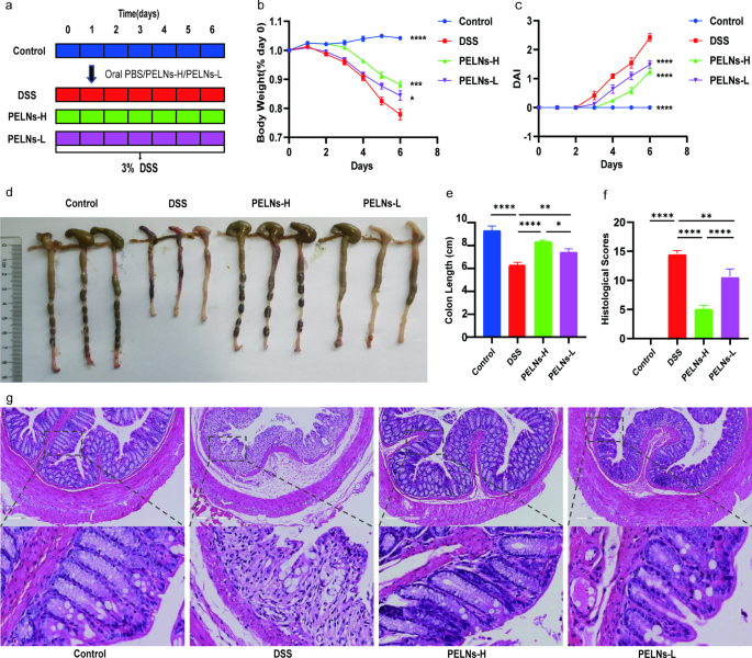
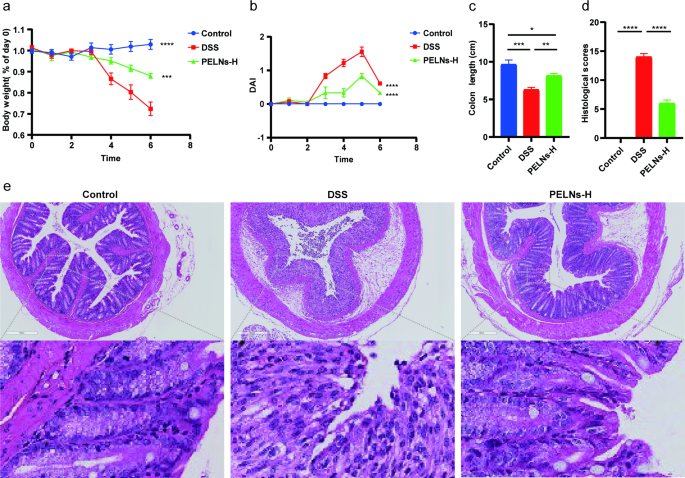
To research the anti-inflammatory results of PELNs, mice have been divided into 4 teams: a wholesome management group, a DSS management group, a PELNs-L group, and a PELNs-H group, as described in Fig. 1a. All through the experiment, the physique weight of mice within the wholesome management group was regularly elevated, whereas a big lower was noticed within the DSS management group (Fig. 1b). Nonetheless, therapy with PELNs confirmed potential in stopping weight reduction in comparison with the DSS management group (Fig. 1b). Notably, PELNs-H gave the impression to be simpler in stopping weight reduction in comparison with PELNs-L (Fig. 1b). Equally, PELNs performed an important position in lowering the illness exercise index (DAI) (Fig. 1c). The PELNs teams exhibited considerably decrease DAI in comparison with the DSS management group (Fig. 1c), and the DAI within the PELNs teams was corresponding to that within the wholesome management group (Fig. 1c). Moreover, PELNs considerably prevented colon shortening (Fig. 1d and e), because the colon size of the PELNs teams was longer in comparison with the DSS management group (Fig. 1d and e). Furthermore, the colon size within the PELNs-H group was barely longer than that within the PELNs-L group, and the fecal situation and mucosa of the colon within the PELNs-H group appeared higher than that within the PELNs-L group, though nonetheless shorter than within the wholesome management group (Fig. 1d and e). Histological evaluation, together with hematoxylin-eosin staining (H&E) and histological scores, additional confirmed that PELNs decreased the buildup of immune cells and harm to the colonic epithelial barrier (Fig. 1f and g). Curiously, the histological scores have been considerably decrease within the PELNs-H group in comparison with the PELNs-L group (Fig. 1g), indicating that PELNs-H had a stronger anti-inflammatory therapeutic impact than PELNs-L within the therapy of DSS-induced colitis in mice.
Oral administration of PELNs protects mice in opposition to DSS-induced colitis. a, Protocol for DSS-induced colitis and PELNs administration; b, Modifications of physique weight over time, normalized to the proportion of the day-zero physique weight; c, Illness exercise index (DAI); d, e, Colon size; f, Histological scores; g, H&E-stained colon sections. *P < 0.05, **P < 0.01, ***P < 0.001 ****P < 0.0001
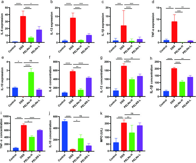
It’s identified that pro-inflammatory cytokines play an important position in intestinal irritation. In colon samples, the degrees of pro-inflammatory cytokines (IL-6, IL-12, IL-1β, and TNF-α), have been considerably elevated within the DSS management group in comparison with the wholesome management group, as decided by qRT-PCR (Fig. 2a, b, c and d). Nonetheless, therapy with PELNs remarkably inhibited the expression of those pro-inflammatory cytokines (Fig. 2a, b, c and d). Conversely, IL-10, an anti-inflammatory cytokine, exhibited a substantial lower within the DSS management group in comparison with the wholesome management group, whereas its ranges have been considerably elevated within the PELNs teams (Fig. 2e).
Professional-inflammatory cytokines and anti inflammatory cytokine expression profiles. a, b, c, d, e, qRT-PCR detecting the degrees of IL-6, IL-12, IL-1β, TNF-α and IL-10 in colonic samples; f, g, h, i, j, ok, ELISA testing the expression profiles of IL-6, IL-12, IL-1β, TNF-α, IL-10 and MPO in blood serum. *P < 0.05, **P < 0.01, ***P < 0.001 ****P < 0.0001
Moreover, an ELISA carried out on blood samples demonstrated a big enhance within the secretion ranges of pro-inflammatory cytokines (IL-6, IL-12, IL-1β, and TNF-α), in addition to myeloperoxidase (MPO), a marker related to neutrophils within the DSS management group in comparison with the wholesome management group (Fig. 2f g, 2 h, 2i, and 2ok). Nonetheless, therapy with PELNs considerably decreased the secretion ranges of those pro-inflammatory cytokines and MPO (Fig. 2f g, 2 h, 2i, and 2ok). Equally, the secretion stage of IL-10 was measured and revealed that PELNs promoted the secretion of IL-10 (Fig. 2j). Notably, PELNs-H exhibited higher efficiency in reducing pro-inflammatory cytokines and growing anti-inflammatory cytokines in comparison with PELNs-L within the therapy of mouse colitis.
Oral administration of PELNs protects IL-10−/− mice acute colitis
To additional assess the anti-inflammatory results of PELNs in an acute colitis mannequin, IL-10−/− mice have been fed and divided into three teams: wholesome management group, DSS management group, and PELNs-H group. Surprisingly, IL-10−/− mice handled with PELNs-H did not develop acute colitis. PELNs-H therapy successfully prevented physique weight reduction, DAI, colon shortening, decreased histological scores and immune cell infiltration in comparison with IL-10−/− mice not handled with PELNs-H (Fig. 3a, b, c and d, and 3e).
Furthermore, qRT-PCR and ELISA outcomes revealed that oral administration of PELNs-H successfully suppressed the expression of pro-inflammatory cytokines (IL-6, IL-12, IL-1β, and TNF-α), in addition to MPO focus in each colon tissues and blood samples of IL-10−/− mice with acute colitis (Determine S2). These findings highlighted the potent anti-inflammatory properties of PELNs-H in mitigating colitis signs within the IL-10−/− mouse mannequin.
In vivo distribution of PELNs
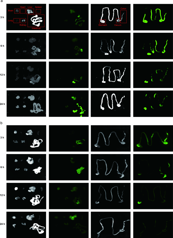
The buildup of oral nanotherapeutics within the desired colon is essential for exerting therapeutic effectiveness in opposition to UC. To be able to decide the biodistribution of PELNs after oral administration, mice with UC have been gavaged with IRDye 800CW-labeled PELNs (100 mg/g) for 3, 6, 12, and 24 h. Close to-infrared imaging revealed that IRDye 800CW-labeled PELNs have been visibly current within the colon at 3 h, and their presence was regularly elevated at 6 h in colitis mice. Nonetheless, the fluorescent indicators detected within the colon have been steadily decreased by 24 h (Fig. 4a). Notably, low fluorescence indicators have been detected within the colon when PELNs have been administered to wholesome mice (Fig. 4b). Subsequently, the buildup of fluorescent indicators in very important organs, corresponding to the guts, liver, spleen, lung, and kidney, was examined. It was discovered that only a few fluorescent indicators have been detected in these organs in mice with colitis handled with PELNs and well being management at 3, 6, 12, and 24 h (Fig. 4a and b). This discovering indicated that PELNs selectively focused the infected colon within the therapy of colitis in mice.
PELNs distinctly altered the variety of the intestinal microflora
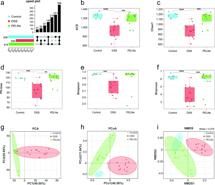
The profiles of intestinal microbiota have been analyzed utilizing 16 S rDNA sequencing. The abundance of intestine microbiota was assessed by calculating the variety of OTUs, which was considerably elevated after PELNs therapy in comparison with colitis mice, as depicted in Fig. 5a. To additional consider the impact of PELNs on α-diversity, 5 range indices have been examined: ACE (Fig. 5b), Chao1 (Fig. 5c), PD-tree (Fig. 5d), Simpson (Fig. 5e), and Shannon (Fig. 5f). DSS-induced colitis led to a big lower in α-diversity as indicated by all 5 indices (ACE, P = 0.00002; Chao1, P = 0.000016; PD-tree, P = 0.000171; Simpson, P = 0.000257; Shannon, P = 0.000015). Nonetheless, therapy with PELNs considerably elevated α-diversity, as mirrored by the upregulation of the ACE (P = 0.000775), Chao1 (P = 0.000707), PD-tree (P = 0.001010), Simpson (P = 0.107145), and Shannon (P = 0.003565) indices. β-diversity was assessed to look at the variations between the intestine microbiota of various teams utilizing PCA (Fig. 5g), PCoA (Fig. 5h), and NMDS (Fig. 5i). Constant outcomes have been noticed throughout all three strategies. The NMDS, PCA (PCA1 + PCA2 = 79.44 > 50%), and PCoA (PCoA1 + PCoA2 = 52.40 > 50%) analyses confirmed that PELNs considerably altered the microbial construction of colitis mice, bringing it nearer to the wholesome management group. Moreover, the stress worth of the NMDS evaluation was 0.078, indicating a superb match to the information. Total, these findings urged that PELNs therapy modulated the composition and construction of the intestinal microbiota in colitis mice, resulting in elevated α-diversity and restoration of the microbial construction in the direction of a more healthy state.
PELNs alters range of intestine microbiota in mice fashions of colitis. a, Upset Polt of OTUs; b, ACE index; c, Chao 1 index; d, PD-tree index; e, Simpson index; f, Shannon index; g, PCA (Principal Part Evaluation); h, PCoA (principal co-ordinate evaluation). Sum of PCoA1 and PCoA2 greater than 50%, indicating vital distinction; i, NMDS (Non-metric Multi-Dimensional Scaling). Stress worth lower than 0.1, indicating vital distinction. **P < 0.01, ***P < 0.001, ****P < 0.0001
PELNs enhance DSS induced adjustments in dominant microbiota and mediated vital adjustments in microbial construction
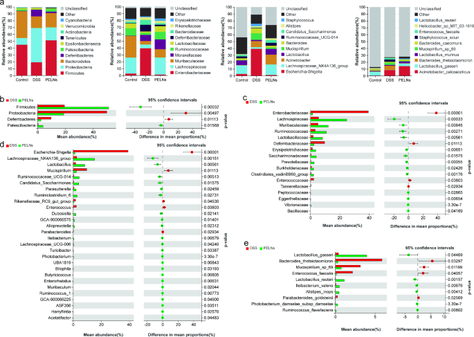
In Fig. 6a, the relative abundance of sure bacterial taxa was considerably totally different between the mice handled with PELNs and the colitis group. Firmicutes (51.26% vs. 19.36%, P = 0.000141), Patescibacteria (3.92% vs. 0.41%, P = 0.006606), Tenericutes (0.92% vs. 0.10%, P = 0.089697), Actinobacteria (0.46% vs. 0.13%, P = 0.071755), Lachnospiraceae (24.75% vs. 5.53%, P = 0.000056), Ruminococcaceae (10.81% vs. 3.18%, P = 0.001079), Muribaculaceae (12.08% vs. 5.09%, P = 0.008093), Lactobacillaceae (8.23% vs. 1.81%, P = 0.002318), Moraxellaceae (14.34% vs. 8.82%, P = 0.502718), Erysipelotrichaceae (3.64% vs. 1.32%, P = 0.004203), Lachnospiraceae_NK4A136_group (15.23% vs. 2.53%, P = 0.000263), Lactobacillus (8.22% vs. 1.81%, P = 0.002315), Acinetobacter (14.34% vs. 8.82%, P = 0.502718), Ruminococcaceae_UCG-014 (4.30% vs. 0.51%, P = 0.001301), Candidatus_Saccharimonas (3.92% vs. 0.41%, P = 0.006644), Alistipes (2.61% vs. 2.02%, P = 0.514264), Acinetobacter_calcoaceticus (14.11% vs. 8.43%, P = 0.484667), Lactobacillus_gasseri (3.95% vs. 0.40%, P = 0.027309), Lactobacillus_murinus (2.80% vs. 1.30%, P = 0.177553), Lactobacillus_reuteri (1.12% vs. 0.03%, P = 0.000201), and Bacteroides_caecimuris (1.82% vs. 1.20%, P = 0.531045) have been notably elevated within the PELNs-treated mice. Conversely, the abundances of Proteobacteria (18.79% vs. 49.81%, P = 0.004250), Deferribacteres (2.28% vs. 9.67%, P = 0.010716), Epsilonbacteraeota (0.76% vs. 2.15%, P = 0.188453), Verrucomicrobia (0.10% vs. 0.42%, P = 0.221802), Cyanobacteria (0.05% vs. 0.09%, P = 0.253980), Enterobacteriaceae (1.37% vs. 39.56%, P < 0.0001), Deferribacteraceae (2.28% vs. 9.67%, P = 0.010716), Bacteroidaceae (2.31% vs. 7.14%, P = 0.070016), Rikenellaceae (3.19% vs. 3.83%, P = 0.631456), Escherichia-Shigella (1.27% vs. 39.13%, P < 0.0001), Mucispirillum (2.28% vs. 9.67%, P0.010716=), Bacteroides (2.31% vs. 7.14%, P = 0.070016), Staphylococcus (0.17% vs. 3.67%, P = 0.164005), Bacteroides_thetaiotaomicron (0.31% vs. 5.87%, P = 0.029962), Staphylococcus_sciuri (0.12% vs. 3.61%, P = 0.162046), Mucispirillum_sp_69 (0.75% vs. 3.15%, P = 0.011562), and Enterococcus_faecalis (0.13% vs. 2.17%, P = 0.037956) have been decreased.
PELNs alters intestine microbiota buildings throughout totally different ranges in mice with colitis. a, Stacked bar plot depicts the construction of intestine microbiota in every group of mice. Left to proper: phylum, household, genus, and species; b-e, Welch’s t check evaluation of PELNs mediated differential microbial adjustments on the phylum, household, genus, and species stage
Welch’s t-test was employed to look at the microbial adjustments induced by PELNs. The evaluation revealed a better abundance of Firmicutes and Patescibacteria (Fig. 6b), whereas Proteobacteria and Deferribacteres exhibited decreased abundance. Notably, the relative proportion of a number of households (Fig. 6c), together with Lachnospiraceae, Ruminococcaceae, Muribaculaceae, Lactobacillaceae, Saccharimonadaceae, Erysipelotrichaceae, Prevotellaceae, Clostridiales_vadinBB60_group, Burkholderiaceae, Vibrionaceae, Eggerthellaceae, Peptococcaceae, and Bacillaceae, confirmed a exceptional enhance. Alternatively, the abundances of Enterobacteriaceae, Deferribacteraceae, Enterococcaceae, and Tannerellaceae have been decreased in comparison with the colitis group. Moreover, on the genus stage (Fig. 6d), the PELNs group exhibited increased relative abundances of Lachnospiraceae_NK4A136_group, Lactobacillus, Ruminococcaceae_UCG-014, Candidatus_Saccharimonas, Ruminiclostridium_6, Dubosiella, Parasutterella, Alloprevotella, GCA-900,066,575, Ileibacterium, Photobacterium, Turicibacter, Lachnospiraceae_UCG-006, Ruminococcus_1, Bilophila, Enterorhabdus, Butyricicoccus, ASF356, UBA1819, Harryflintia, GCA-900,066,225, Muribaculum, and Acetatifactor, whereas Escherichia-Shigella, Mucispirillum, Enterococcus, Rikenellaceae_RC9_gut_group, and Parabacteroides exhibited decrease abundances. On the species stage (Fig. 6e), the relative proportions of Lactobacillus_gasseri, Lactobacillus_reuteri, Alistipes_inops, Ileibacterium_valens, Photobacterium_damselae_subsp_damselae, and Ruminococcus_flavefaciens have been drastically elevated by PELNs, whereas Bacteroides_thetaiotaomicron, Mucispirillum_sp_69, Enterococcus_faecalis, and Parabacteroides_goldsteinii confirmed decreased proportions. (The Welch’s t-test outcomes evaluating the management group to the DSS group will be discovered within the supplementary supplies, Determine S3).
PELNs-Lactobacillus reuters-indole-derivatives axis induces the differentiation of DP CD4+CD8+T cells
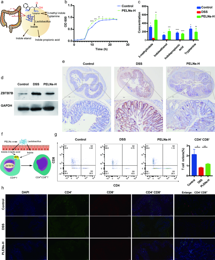
It has been found that Lactobacillus reuteri had the flexibility to metabolize tryptophan (L-Trp) into indole derivatives. These derivatives might activate the aryl hydrocarbon receptor (AhR) in typical CD4+ T cells, resulting in the downregulation of Zbtb7b expression and the reprogramming of typical CD4+ T cells into DP CD4+CD8+ T cells (Fig. 7a and f) [37]. To evaluate the expansion habits of Lactobacillus reuteri, OD600 measurements have been carried out in vitro with and with out PELNs. The outcomes demonstrated that whereas the expansion of isolates was comparable between the PELNs group and the management group throughout the stationary and delay durations, the expansion of isolates was noticeably quicker within the logarithmic section within the PELNs group in comparison with the management group (Fig. 7b). Moreover, PELNs therapy considerably elevated the degrees of indole derivatives, together with 3-methylindole, indoleethanol, indolepropionic acid and tryptamine, in fecal samples as in comparison with the DSS group (Fig. 7c and Determine S4).
PELNs inducing the differentiation of DP CD4+CD8+T cells. a, Schematic illustration of mechanisms for Lactobacillus reuteri metabolizing tryptophan (L-Trp) into indole derivatives; b, PELNs promote the expansion of Lactobacillus reuteri; c, PELNs therapy considerably elevated the degrees of indole derivatives; d, e, PELNs therapy considerably decreased the degrees of Zbtb7b protein utilizing WB and IHC assay; f, Schematic illustration for the derivatives of Lactobacillus reuteri activating the aryl hydrocarbon receptor (AhR) in typical CD4+ T cells, and resulting in the downregulation of Zbtb7b expression and the reprogramming of typical CD4+ T cells into DP CD4+CD8+ T cells; g, Movement cytometry detecting the inhabitants of DP CD4+CD8+ T cells in colonic samples; h, Symbolize immunofluorescence exhibiting the inhabitants of DP CD4+CD8+ T cells in colonic samples. Knowledge are consultant FCM pictures or expressed because the imply ± SEM of every group. *P < 0.05, **P < 0.01
DP CD4+CD8+ T cells are localized within the intestinal epithelial layer and serve a number of capabilities, together with the suppression of intestinal irritation induced by sort 1 helper T (Th1) cells, upkeep of intestine mucosal homeostasis, and inhibition of pro-inflammatory cytokine launch throughout pathogenic infections [37]. It has additionally been noticed that the down-regulation of Zbtb7b in intraepithelial T lymphocytes (IELs) represses CD4+ T cell differentiation whereas selling the differentiation of CD8+ T cells, resulting in the reprogramming of CD4+ T cells into DP CD4+CD8+ T cells [37]. In our earlier research, we’ve demonstrated a big enhance in Zbtb7b expression and a notable lower within the inhabitants of DP CD4+CD8+ T cells in each UC sufferers and UC mouse colon samples [36]. Due to this fact, we aimed to research the impression of oral administration of PELNs on the differentiation of DP CD4+CD8+ T cells in mice with colitis. Strikingly, we noticed a big discount within the expression of Zbtb7b on the protein stage within the PELNs group, as demonstrated by WB evaluation and IHC assay (Fig. 7d and e). Moreover, the inhabitants of DP CD4+CD8+ T cells was considerably increased within the PELNs group in comparison with the DSS management group, as decided by FCM (1.1% vs. 0.75%, P < 0.01, Fig. 7g). Nonetheless, there was a big distinction within the inhabitants of DP CD4+CD8+ T cells between the PELNs group and the wholesome group (1.1% vs. 2.45%, P < 0.05, Fig. 7g). Equally, IF evaluation revealed a marked enhance within the inhabitants of DP CD4+CD8+ T cells within the PELNs group in comparison with the DSS management group (Fig. 7h). Moreover, the inhabitants of DP CD4+CD8+ T cells within the PELNs group was corresponding to that of the wholesome group (Fig. 7h).
Biosafety of orally administered PELNs
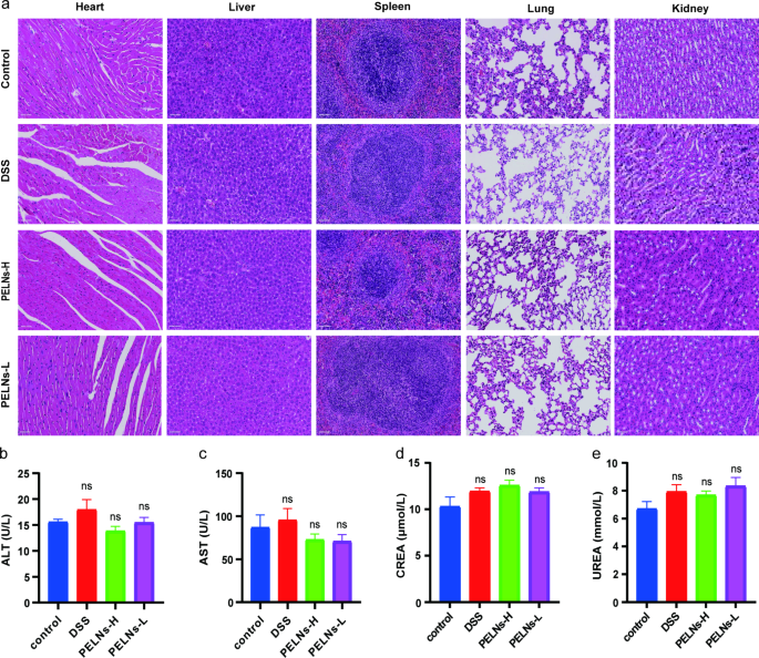
To evaluate the security of orally administered PELNs, blood serum samples and very important organs (coronary heart, liver, spleen, lung, and kidney) have been collected from mice that obtained day by day doses of fifty mg/g and 100 mg/g PELNs for five days. Histological evaluation of the very important organs utilizing H&E staining revealed no evident abnormalities or indicators of organ harm within the PELNs-administered group (Fig. 8a). Moreover, blood cell depend, cardiac enzymes, glutamic pyruvic transaminase (ALT), aspartate aminotransferase (AST), serum creatinine (CREA), and serum urea (UREA) didn’t present vital adjustments between the traditional management group, DSS group, and PELNs-administered group (Fig. 8b, c, d and e).


