Construction and composition traits of PLGA and MBR gels
The preparation technique of implants of minimally invasive carriers must be environment friendly and handy. To assemble the bioactive MBRs, PLGA/NMP answer (PLGA gels) was first obtained, as proven in Scheme 1 A. The liquid PLGA gels have been synthesized by shaking PLGA particles with NMP answer. PLGA, a compound authorised by the U.S. Meals and Drug Administration (FDA) for medical use, has been used as a sustained-release drug service, synthetic catheter, and tissue engineering scaffold materials in a number of therapy fields, which suggests its additional potential in tumor therapeutics [25, 26]. The artificial PLGA gels with a very good liquid fluidity may be equally loaded with a number of practical particles, resembling Fe3O4, GOx and MgCO3. After the dispersion of small particles of Fe3O4, GOx and MgCO3 (Determine S1A-C) into the composite PLGA gels, the MBRs have been simply obtained as black gels (Determine S2). Notably, stable PLGA has a porous construction (Determine S1D), which suggests the flexibility to launch hydrosoluble GOx into its environment. As proven in Determine S3, MBRs remained a homogeneous combination even after 8 h, implying that the Fe3O4, GOx and MgCO3 particles may obtain steady distribution within the PLGA gel. Importantly, the superb stability of MBRs endows operators with adequate time for hydrogel implantation.
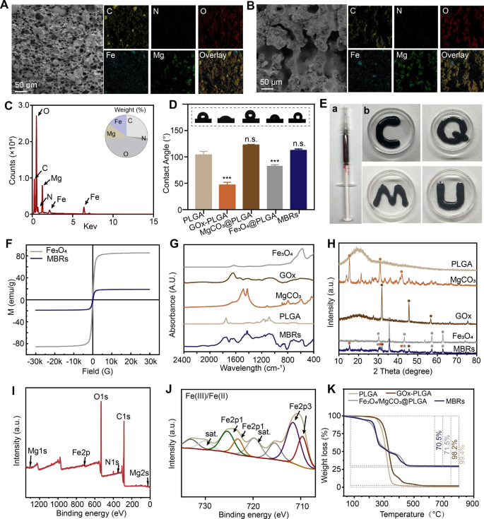
The SEM pictures and corresponding mapping pictures additional verified a uniform porous construction with a diameter lower than 50 μm and homogeneous distributions of C, N, O, Fe and Mg for many MBRs (Fig. 1A). Furthermore, after AMF publicity, the inner morphology of the MBRs exhibited an elevated porous “bridge-like” construction with favorable steady component dispersion (Fig. 1B). The corresponding component quantitative end result by vitality spectrum is proven in Fig. 1C. As well as, the porosity of the MBRs was 40.3 ± 2.3% with out AMF software, whereas it elevated to 68.9 ± 4.2% after AMF publicity, indicating that GOx might be launched into tumors by AMF triggering. As proven in Fig. 1D, after liquid‒stable transformation, the MBRs displayed no distinction from PLGA by way of hydrophilicity, which indicated that there was no impact on cell adhesion. For irregular bone defect restore biomaterials utilized in a minimally invasive technique, good injectability and form adaptability are very important. The liquid type of MBRs have been taken into a typical syringe (Fig. 1E-a). It was discovered that the MBRs may freely move by way of the syringe attributable to their low viscosity and kind numerous irregular floor profiles (Fig. 1E-b). To be used in magnetic hyperthermia, a passable magnetic property is important [27, 28]. Because the lengthy and slim formed hysteresis curve (Fig. 1F) reveals, the MBRs are smooth magnetic ferrite and have a low coercive pressure and residual magnetization worth (saturation magnetization: 18.92 emu/g), much like these of pure Fe3O4 nanoparticles (saturation magnetization: 85.52 emu/g). As a smooth magnetic materials with comparatively low hysteresis, loss properties and coercive pressure, MBRs will be simply triggered by an AMF as a result of the coercive pressure is smaller than that of the AMF [29]. ICP-OES quantitative measurement proven that the iron concentrations of MBRs within the first batch with and with out heating have been 108.196 ± 1.097 mg/g and 108.516 ± 0.6 mg/g, respectively, indicating no vital distinction between the 2 teams (p > 0.05) and illustrating that no Fe3O4 nanoparticles escaped from the MBRs after heating.
Fabrication and characterization of varied types of MBRs: Fe3O4/GOx/MgCO3@PLGA gels. SEM pictures and corresponding mapping pictures of MBRs with out (A) and with (B) AMF publicity. (C) Vitality spectrum of MBRs (insert determine displaying weight% evaluation of components). (D) WCA check outcomes of various teams (insert: optical pictures). (E) Digital pictures of the injectable MBRs by way of a typical 1 ml syringe and consultant pictures of various kinds. (F) Magnetic hysteresis loop of MBRs and Fe3O4 nanoparticles. (G) FTIR spectra of PLGA, MgCO3, GOx, Fe3O4 and MBRs. (H) XRD spectra of PLGA, MgCO3, GOx, Fe3O4 and MBRs. (I) XPS spectra of MBRs. (J) Fe 2p XPS of MBRs. (Okay) TGA curves of PLGA, GOx-PLGA, Fe3O4/MgCO3@PLGA and MBRs.
To additional decide the composition and chemical building elemental standing of the MBRs, a number of check analyses of the as-prepared stable samples have been carried out. The FTIR spectrum of MBRs indicated the attribute absorption peaks of MgCO3 and GOx, confirming the presence of MgCO3 and GOx molecules (Fig. 1G). Furthermore, the outcomes of the X-ray diffraction (XRD) assay (Fig. 1H) steered that the chemical construction of the included Fe3O4 and MgCO3 wouldn’t be modified by the preparation and liquid‒stable section transition technique of the MBRs. Notably, the presence of the weather C, N, O, Mg, and Fe was confirmed by X-ray photoelectron spectroscopy (XPS) evaluation related to C 1s, N 1s, O 1s, Mg 2s, and Fe 2p, respectively (Fig. 1I). The C 1s spectrum of the C-O peak at 532.5 eV (Determine S4) is in distinction with the C 1s spectrum of the MBRs, which reveals a brand new increased C-H peak at 284.6 eV (Determine S5), attributed to the C-H of the GOx particles. As well as, the attribute Mg 1s peak at 1034.5 eV of the shakeup satellite tv for pc peaks similar to that of Mg2+ was noticed from the high-resolution Mg 1p XPS spectrum of the MBRs (Determine S6). In comparison with the stable type of PLGA, the emerged attribute Fe 2p peaks at 710.5 eV (Fe 2p3) and 725.9 eV (Pt 2p1) have been attributed to that of Fe2+ and Fe3+, which was attributable to the modification of the Fe(III)/Fe(II) prodrug (Fig. 1J). The presence of Mg and Fe indicated excessive potential for triggering magnetic hyperthermia and Mg-related bone restore. Moreover, after the heating course of (from 37 ℃ to 800 ℃), the residual composition within the numerous scaffolds was estimated, as proven in Fig. 1Okay. The MBRs confirmed good thermal stability underneath heating situations, and no apparent thermal decomposition of the fabric was noticed within the magnetothermal working temperature vary (< 50 ℃). The numerous weight reduction noticed for Fe3O4/MgCO3@PLGA and the MBRs upon heating was ascribed to the thermal decomposition of MgCO3. In contrast with Fe3O4/MgCO3@PLGA, the roughly 1% weight lack of the MBRs was in line with the loading dose of GOx. Collectively, these good performances of the MBRs indicated that MgCO3, Fe3O4 and GOX have been efficiently loaded into the PLGA gels.
Magnetic hyperthermia traits of MBRs
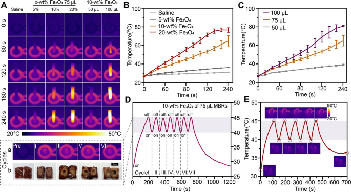
To realize controllable ranges of heating temperatures and gentle magnetothermal results in vivo, the optimum Fe3O4 mass ratio must be precisely evaluated. The outcomes of magnetothermal exams indicated that the presence of MBRs may shortly and successfully enhance the temperature of the environment in response to the infrared thermography pictures (Fig. 2A) and corresponding temperature curves (Fig. 2B). The saline answer with out magnetic supplies confirmed no vital temperature variations. For Fe3O4-MBRs loaded with numerous mass ratios (10%, 20%), the hyperthermia temperature simply reached over 45 °C, whereas the 5% Fe3O4-MBRs didn’t exhibit this efficiency. For the 20% Fe3O4-MBRs, the ultimate temperature of the encircling water tub was over 80 °C. As quantity of 10% Fe3O4-MBRs elevated, the magnetothermal impact improved (Fig. 2C). Nonetheless, for 100 µL of 10% Fe3O4-MBRs, the temperature management property was not ideally suited attributable to its steep temperature curve, which exceeded 80 °C and was not appropriate for gentle magnetothermal remedy. Therefore, 75 µL of 10% Fe3O4-MBRs was optimum for additional experiments. To simulate the intratumor therapeutic vary to keep away from injuring surrounding wholesome tissue, the ex vitro bovine liver was used to judge the therapy impact.
Delicate magnetothermal efficiency of varied types of MBRs-Fe3O4/GOx/MgCO3@PLGA gels in vitro and in vivo. (A) In vitro infrared thermal pictures of Fe3O4/MgCO3@GOx-PLGA with completely different volumes and numerous mass fractions of Fe3O4 nanoparticles. (B) Corresponding quantitative temperature curves of saline and MBRs (included with completely different mass fractions of Fe3O4 nanoparticles). (C) Corresponding quantitative temperature curves of 10% Fe3O4-MBRs with numerous volumes. (D) Quantitative temperature curves of 75 µL 10% Fe3O4-MBRs for “on-off” mannequin magnetothermal in vitro bovine liver therapy with corresponding infrared thermal pictures and digital pictures of a number of cycles. (E) Temperature-time curve of 143B tumor-bearing mice after intratumoral injection of MBRs and AMF publicity for 5 on-off cycles and the corresponding infrared thermal pictures
Temperatures within the vary of 40 ~ 45 °C cannot solely eradicate most cancers cells immediately with little harm to surrounding regular tissue but in addition induce mixture remedy, particularly for GOx as an enzyme compound retaining protein exercise [30, 31]. Therefore, we tried to use an “on-off” cyclic heating mannequin that maintained the goal temperature in an efficient vary [32]. As Fig. 2D-a reveals, the 75 µL of 10% Fe3O4-MBRs offered good magnetic thermal stability through the thermal course of, and the temperature of the middle of the bovine liver persistently and mildly elevated. After a number of cycles of magnetic thermal heating, the morphology of the bovine livers and the ablation vary have been measured, displaying livers with pale tissue and apparent coagulative necrosis underneath a microscope after therapy (Fig. 2D-b). For the 5 heating cycles, the therapeutic vary was roughly 1 cm, which is the perfect match to be used in tumors (Determine S7). Furthermore, the publicity period was chosen primarily based on the tumor quantity, which probably presents a person therapy scheme for sufferers with numerous tumor sizes by various the publicity period and implanted content material of MBRs. The “on-off” cyclic heating technique can also be applicable for software in vivo (Fig. 2E). In 143B tumor-bearing mice, the MBRs additionally exhibited good magnetothermal stability through the heating course of. Consequently, 75 µL of 10% Fe3O4-MBRs was chosen on this work because the optimum gentle MTT situation for triggering GOx launch, and it’s a rational alternative for subsequent MTT and hunger remedy.
Delicate hyperthermia-triggered GOx launch for inducing hunger remedy
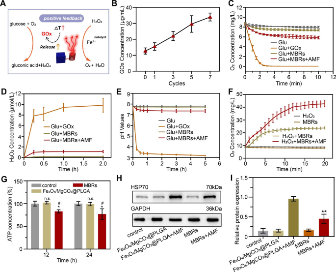
To discover the mechanism by which gentle hyperthermia triggers GOx launch to induce hunger remedy, the GOx-mediated response course of and the mutual selling impact have to be additional verified. As anticipated, gentle magnetic hyperthermia enhanced the discharge of GOx by the MBRs to speed up the transformation of glucose (Glu) into gluconic acid and H2O2, and H2O2 may additional rework into O2 and H2O, particularly within the thermal surroundings (Fig. 3A). When GOx is used as an antitumor drug in biomaterials, we anticipate it to play a tumor killing function and scale back the affect on surrounding regular cells by way of managed launch. In line with the usual curve of the GOx answer (Determine S8), the cumulative launch focus of GOx from the MBR + AMF group was a lot increased than that from the MBR group (Determine S9), which signifies that triggered GOx launch was achieved by the AMF. Moreover, the quantity of GOx launch was positively associated to “on-off” therapy cycles (Fig. 3B), which illustrated that MTT was a driver of MBRs for the GOx response. The oxygen focus was measured by a transportable dissolved oxygen meter to look at the GOx-mediated oxygen consumption potential [33]. In line with the time-dependent curves in Fig. 3C, the focus of oxygen within the Glu + GOx group quickly decreased because the response continued and reached a plateau in 3 min, whereas the Glu group displayed virtually no response. This end result indicated that soluble oxygen might be produced by way of GOx-mediated glucose oxidization. As well as, as a result of GOx was encapsulated within the MBRs, the oxygen content material of the Glu + MBRs group decreased extra slowly than that of the Glu + GOx group, i.e., free GOx answer. Furthermore, because of the launch of activated GOx from the MBRs, the oxygen focus within the Glu + MBRs + AMF group decreased extra shortly than that within the Glu + MBRs group, which implied that MTT is a optimistic promoter of the GOx-mediated hunger response. Nonetheless, in contrast with the Glu + GOx group, the oxygen focus within the Glu + MBRs + AMF group decreased slowly, demonstrating the reoxygenation capability of MBRs + AMF by thermal decomposition or by Fe3+ catalyzing H2O2 to O2 and indicating that this technique may speed up substrate consumption for propulsive reactions.
Delicate hyperthermia-triggered GOx launch to induce hunger remedy. (A) Schematic illustration of the mechanism of the gentle hyperthermia-triggered GOx enzymatic response. (B) Launch of GOx from MBRs after AMF publicity. (C) Oxygen focus adjustments over time in numerous options of glucose, GOx + glucose, MBRs + glucose, and MBRs + AMF + glucose. (D) Modifications in H2O2 focus ensuing from reactions with GOx + glucose, MBRs + glucose, and MBRs + AMF + glucose at numerous time factors. (E) Modifications in pH ensuing from the response between glucose, GOx + glucose, MBRs + glucose, and MBRs + AMF + glucose at numerous time factors. (F) Time-dependent curves of oxygen focus for H2O2, MBRs, H2O2 + MBRs, and H2O2 + MBRs + AMF. (G) ATP focus of 143B cells from completely different teams after AMF publicity. (H) Consultant immunoblot outcomes of various protein ranges of HSP70 for every therapy group and (I) corresponding quantitative analyses of the ratios of HSP70/GAPDH. (The information are proven because the means ± SDs, n = 3 per group, n.s. represented no significance and *p < 0.05, **p < 0.01, as compared with the management teams, ##p < 0.001 as compared with Fe3O4/MgCO3@PLGA, respectively.)
The identical ideas have been utilized for detecting substrate manufacturing. The oxygen focus within the Glu + GOx + H2O2 group elevated shortly inside 15 min, whereas negligible adjustments have been detected within the Glu + MBRs + AMF + H2O2 teams (Fig. 3F). Furthermore, with the manufacturing of gluconic acid, the pH continued to lower [34]. Because the hunger response progressed, the H2O2 and gluconic acid concentrations step by step elevated (Fig. 3D and E), which confirmed that the managed launch of GOx was achieved by triggering the MBRs by way of MTT, successfully catalyzing the glucose oxidation response and changing glucose into gluconic acid and H2O2. As Fig. 3F reveals, within the H2O2 + MBR + AMF group, H2O2 decomposed into H2O and O2 underneath the gentle thermal therapy because the response occurred, and Fe3+ decomposed from Fe3O4 underneath the AMF to facilitate H2O2 dissociation. With out AMF triggering, the quantity of H2O2 decomposition markedly decreased. These outcomes additional confirmed that MTT might be helpful for the development of GOx-mediated hunger reactions.
The optimistic suggestions of gentle hyperthermia–hunger mutual synergistic remedy can also contain a thermoresistance remission mechanism. Earlier research have proven that HSPs produced within the warmth response course of could cause tumor warmth resistance, and this pathway could be very depending on ATP as an vitality provide [35, 36]. Due to this fact, delaying ATP manufacturing is predicted to beat the warmth resistance of tumors and improve the therapeutic impact of MTT. As anticipated, a comparatively low ATP focus was detected after therapy with MBRs after AMF irradiation (Fig. 3G), presumably as a result of the hunger response consumes glucose. HSP70 expression in 143B cells underneath completely different therapies was evaluated by Western blot (WB) evaluation. In line with the anticipated outcomes, excessive HSP70 expression was noticed in 143B cells handled with Fe3O4/MgCO3@PLGA after AMF publicity, immediately indicating a defensive warmth shock response by way of thermal remedy (Fig. 3H). The MBRs + AMF therapies considerably decreased the HSP70 expression ranges, and the outcomes have been in line with ATP focus variation, verifying that HSP overexpression might be inhibited by interfering with ATP manufacturing. After the intervention of 143B cells with completely different therapies, the HSP70 ranges have been measured. After Fe3O4/MgCO3@PLGA + AMF therapy, the HSP70 stage elevated 3.82-fold. In distinction, the MBRs + AMF group considerably decreased the relative stage of HSP70 by 1.95-fold (Fig. 3I). Taken collectively, these outcomes counsel that GOx might be an efficient adjuvant for enhancing MBRs-based MTT by downregulating HSP expression.
Efficacy of mutually synergistic gentle hyperthermia–hunger remedy in vitro
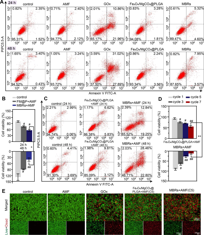
Good biocompatibility and biosafety are stipulations of responsive functionalized implants for in vivo purposes, and the harmlessness of the MBRs with out utilizing an AMF was anticipated. Due to this fact, the biocompatibility and biosafety of the MBRs and their therapeutic results have been examined by cell apoptosis utilizing stream cytometry. For GOx answer with out PLGA gel, the proportion of viable cells confirmed a pointy decline of 62.5% in coculture for 48 h. Nonetheless, the MBRs confirmed excessive biosafety with out AMF publicity when cocultured with cells for twenty-four and 48 h, indicating the harmlessness of the MBRs with PLGA gels. In comparison with the MBRs group, the focus of GOx within the GOx group was increased when GOx was immediately added into the tumor cell tradition system. The extreme GOx quickly consumed glucose within the tradition medium and produced H2O2, inhibiting tumor cell metabolism and rising oxidative stress ranges throughout the tumor cells, in the end killing tumor cells, which could trigger a pointy lower in blood sugar and intrude with regular cell metabolism, and isn’t appropriate for in vivo therapy. Nonetheless, when mixed with AMF triggering, the cell viability declined underneath coincubation with Fe3O4/MgCO3@PLGA and MBRs. In distinction, the Fe3O4/MgCO3@PLGA group’s cytotoxic efficacy was a lot decrease than that of the MBRs teams with the identical therapy and irradiation time because the CCK-8 assay (Fig. 4B) and stream cytometry evaluation (Fig. 4C). The GOx launch triggered by magnetic hyperthermia and the mutually synergistic hyperthermia–starviation remedy impact may clarify this end result.
In vitro gentle hyperthermia-starving mutual synergistic remedy in opposition to 143B cells. (A) Movement cytometry was used to find out and analyze the apoptosis of 143B cells cocultured with PBS, AMF, GOx, Fe3O4/MgCO3@PLGA, and MBRs for twenty-four and 48 h. (B) Cell viability assay and (C) stream cytometry evaluation of 143B cells incubated with PBS, Fe3O4/MgCO3@PLGA and MBRs after AMF publicity for twenty-four and 48 h. (D) Cell viability assay outcomes of 143B cells after numerous cycles of “on-off” mannequin magnetothermal therapy for Fe3O4/MgCO3@PLGA and MBRs teams. (E) Calcein-AM (inexperienced)/PI (purple) staining pictures of 143B cells after numerous therapies (scale bars: 200 μm). (The information are proven because the means ± SDs, n = 3 per group, n.s. represents no significance and #p < 0.05, ##p < 0.01, as compared with the management teams, *p < 0.05 **p < 0.01 as compared between teams, respectively.)
As anticipated, after it was decided that the MBRs have very good magnetic thermal properties and the flexibility to set off GOx launch to induce hunger remedy, their antitumor efficacy underneath AMF publicity was investigated as one other necessary property. As talked about above, because the variety of therapy cycles elevated, the in vitro magnetic hyperthermia impact improved. Cell viability was additional evaluated with a CCK-8 assay, and an analogous end result was obtained. For the MBR group, after seven cycles of AMF publicity, the survival fee of the cells was markedly decreased and virtually converged to zero after 48 h (Fig. 4D). To forestall regular tissue harm, the five-cycle “on-off” technique was deemed appropriate for additional experiments, reaching comparable efficacy to that of in vitro magnetic hyperthermia. The inhibitory impact of the MBRs on 143B cells was visually examined utilizing a calcein-AM/PI staining assay. Stay cells (inexperienced fluorescence staining) and lifeless cells (purple fluorescence staining) have been noticed by fluorescence microscopy. The management and AMF-only teams exhibited no purple fluorescence, indicating that an AMF has no cell killing potential towards 143B cells. Extra pronounced purple fluorescence was noticed within the GOx answer group, indicating that the predominant cell-killing impact of GOx was impartial of AMF triggering, which could be related to stopping unintended damage to regular cells (Fig. 4E). Extra importantly, as Fig. 4E and Determine S10 present, the MBRs included with GOx produced robust purple fluorescence after 5 cycles of therapy, displaying an enhanced however considerably comparable therapeutic impact to that of Fe3O4/MgCO3@PLGA. These outcomes demonstrated that mutual synergistic hyperthermia–hunger remedy had a wonderful impact in opposition to 143B cells.
Antitumor therapeutic efficacy and biosafety of the MBRs
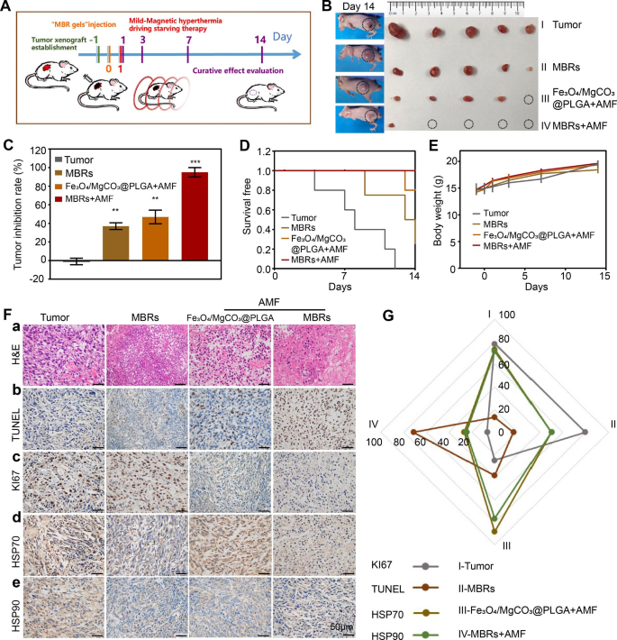
Contemplating the predominant healing impact in vitro and the great stable transformation efficiency of the MBRs in tumor tissues, we anticipate that MBRs may operate as triple-functional magnetic gels for in vivo osteosarcoma remedy. Following therapeutic course of described in Fig. 5A, we established a 143B human osteosarcoma tumor-bearing nude mouse mannequin and stochastically divided the mice into 4 teams: Tumor group (PBS), MBR group (MBRs), Fe3O4/MgCO3@PLGA with AMF publicity group (Fe3O4/MgCO3@PLGA + AMF), and MBRs with AMF publicity group (MBR + AMF). The mice have been injected with completely different gels (75 µL per mouse), adopted by AMF irradiation (5 cycles). As a result of MBRs present very good temperature management, the tumor-site temperature within the Fe3O4/MgCO3@PLGA + AMF group and the MBRs + AMF group was precisely elevated to 40–45 °C through the publicity cycles (Fig. 2E). After therapy with completely different interventions, the amount of the tumors and the physique weights have been calculated at completely different time factors (Fig. 5A). Because the tumor picture (Fig. 5B) and inhibition fee (Fig. 5C) proven, the tumors within the saline-treated group grew quickly (Tumor group-control), with a most tumor quantity over 1 cm3. For therapy with MBRs alone, the tumor progress inhibitory impact was unsatisfactory (inhibition fee: 37 ± 3.6%). This was additionally the case for mice handled with Fe3O4/MgCO3@PLGA plus AMF publicity (inhibition fee: 46.9 ± 7.4%). As a result of the quantity of GOx launched by the MBRs was small, the effectivity of GOx-mediated glucose depletion was low, and utilizing solely magnetic hyperthermia led to residual tumor. Compared, a marked inhibition of tumor measurement was noticed within the MBRs + AMF group (inhibition fee: 95 ± 5%), which is because of the impact of mutually synergistic gentle hyperthermia–hunger mutual synergistic remedy. The morbidity-free survival from every therapy group additional confirmed the above outcomes (Fig. 5D). Furthermore, no vital body-weight variations have been noticed in every group (Fig. 5E), indicating that every practical particle of MBRs had excessive biosafety and biocompatibility for in vivo purposes.
Antitumor therapeutic efficacy of MBRs. (A) Remedy and follow-up routine. (B) Image of the excised 143B tumors 14 days after therapies and the corresponding tumor-bearing mice. (C) Tumor inhibition fee of 143B tumor-bearing mice with the assorted therapies. (D) Morbidity-free survival and (E) physique weight of 143B tumor-bearing mice within the completely different therapy teams. (F) H&E staining (a) and immunohistochemical staining (TUNEL-b, Ki67-c, Hsp70-d and Hsp90-e) within the tumor area of every group. (G) Quantitative evaluation of immunohistochemical staining (Fig. 5-F). (Scale bars: 50 μm). (The information are proven because the means ± SDs, n = 5 per group, n.s. represented no significance and *p < 0.05, **p < 0.01, ***p < 0.001 as compared with the management teams, respectively.)
To additional consider the substantial harm of various teams to tumor cells and the corresponding potential mechanisms, H&E, KI67, and TUNEL staining have been carried out. In H&E staining, the mixture of MBRs with AMF irradiation prompted essentially the most vital large-area cell shrinkage and extreme necrosis (Fig. 5F-a). Solely Fe3O4/MgCO3@PLGA + AMF resulted in reasonable cell necrosis, whereas no apparent cell harm was discovered within the different two teams. The above cell morphology indicated that completely different therapeutic methods produced completely different therapeutic results. Immunohistochemistry (IHC) staining was additionally carried out to judge the apoptotic index and proliferative index after completely different therapies. Essentially the most distinct 143B cell apoptosis, as noticed by TUNEL staining of the nucleus (Fig. 5F-b), was noticed after therapy with MBRs mixed with AMF irradiation, which additionally proven the best inhibitory impact on proliferation, as proven by KI67 staining (Fig. 5F-c).
To additional illustrate the underlying mechanism of synergistic remedy, IHC staining of HSP70 and HSP90 was used to research the inhibitory impact of MBRs-mediated mutual synergistic hyperthermia–hunger remedy on these heat-resistant proteins. As proven in Fig. 5F-d and e, the expression ranges of HSP70 and HSP90 within the Fe3O4/MgCO3@PLGA + AMF group have been a lot increased within the cytoplasm, as indicated by darkish brown staining, than these within the MBRs group, revealing that the expression of HSP70 and HSP90 could also be induced by magnetic hyperthermia. Nonetheless, the IHC staining depth outcomes of the MBR + AMF group have been clearly decrease than these of the Fe3O4/MgCO3@PLGA + AMF group as a result of magnetic hyperthermia triggered the GOx release-mediated hunger impact by way of decreased ATP era to inhibit HSP expression. Furthermore, the IHC intensities of HSP70 and HSP90 have been analyzed, and the outcomes have been the identical (Fig. 5G).
To judge the potential of medical MBRs transformation, the biocompatibility of the MBRs was evaluated with regard to their potential use as therapeutic brokers. First, the H&E staining diagram (Determine S11) and organ weight (Determine S12) of the main organs (together with the center, liver, spleen, lung and kidney) of the MBRs-treated mice confirmed no apparent abnormalities or tissue harm, indicating the negligible toxicity of the MBRs to the primary organs. Furthermore, blood samples have been collected from mice after numerous therapies to judge the corresponding systemic toxicity (Determine S13). In contrast with these within the management group, not one of the hematological parameters within the MBR group have been considerably modified at 7 and 28 days after injection. These outcomes steered that MBRs are extremely biocompatible as promising therapeutic brokers for mutual synergistic hyperthermia–hunger remedy in opposition to 143B OS in vivo.
MBRs accelerated osteogenesis in vitro and in vivo
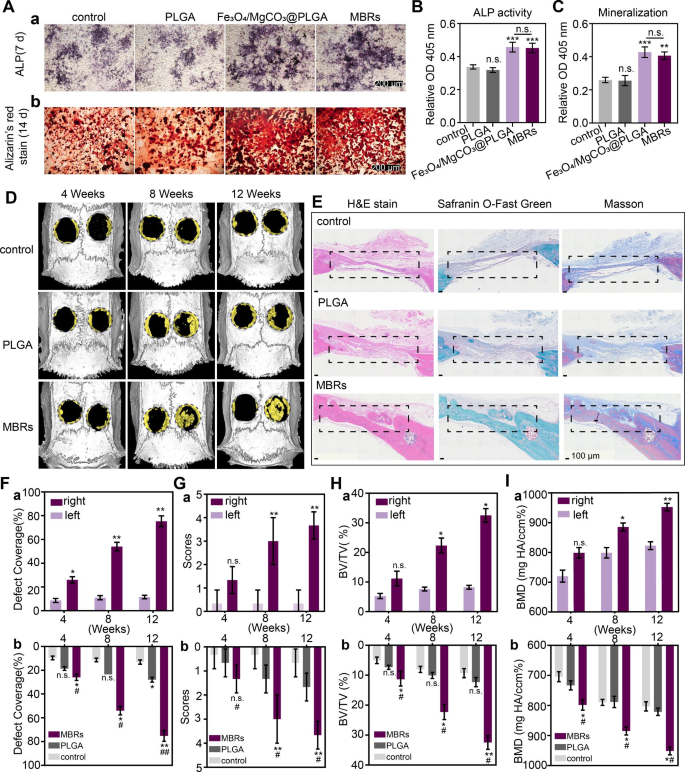
Contemplating the bioactive results of magnesium ions, resembling therapeutic osteoinductive features and enhancing matrix mineralization, MgCO3 was loaded into PLGA gels as a Mg2+ supply to acquire Fe3O4/GOx/MgCO3@PLGAgels (MBRs). Moreover, the osteogenic peculiarities of MBRs have been evaluated in vitro and in vivo. ALP staining on day 7 implied the distinguished osteogenic differentiation of iMEFs within the Fe3O4/MgCO3-PLGA group and MBRs group in contrast with the management group and PLGA group (Fig. 6A-a). That is almost definitely as a result of the Fe3O4/MgCO3-PLGA group and the MBRs group each exhibited sustainable Mg2+ launch. Moreover, quantitative evaluation proven that ALP exercise was additionally considerably increased than that within the different teams, indicating that MBRs promoted the osteogenic differentiation of iMEFs, whereas PLGA gels alone didn’t present this property (Fig. 6B). Along with the improved ALP manufacturing, ARS staining was used to immediately visualize mineralization (Fig. 6A-b), which fashioned on the late stage of osteogenic differentiation and was assessed by quantitative evaluation (Fig. 6C), revealing an apparent enhance in mineral deposition induced by the MBRs. Therefore, MBRs have the best capability to advertise the osteogenic differentiation of iMEFs in vitro.
Analysis of the MBRs potential for bone regeneration in vitro and in vivo. (BA) ALP staining and alizarin purple staining (ARS) of PBS, PLGA, Fe3O4/MgCO3@PLGA and MBRs. Quantitative evaluation of (B) ALP exercise and (C) ARS staining. (D) Reconstructed 3D micro-CT pictures of rat crania with the handled defects (left: self-blank management, proper: therapy with completely different teams). (E) Histological analysis of bone defect regeneration by H&E, safranin O-fast inexperienced and Masson’s trichrome staining (the restore space is labeled with rectangular containers, scale bars: 100 μm). Quantitative evaluation of bone defect regeneration as decided by (F) the defect protection %, (G) the rating of bone defect restoration, (H) BV/TV% and (I) BMD: (a) throughout the MBRs group and (b) among the many three teams. (The information are proven because the means ± SDs, n = 3 per group, n.s. represented no significance and *p < 0.05, **p < 0.01 as compared with the management teams, # p < 0.05 ## p < 0.01 as compared with PLGA teams, respectively
As designed, MBRs with good injectability and form adaptability have been injected into established cranial defects and stabilized after section transformation. The appropriate facet was the therapy group, and the left facet was used for the self-control examine. After twelve weeks of implantation, extra freshly generated osteogenic tissue was noticed within the MBRs group than within the PLGA group, as decided by micro-CT (Fig. 6D); nevertheless, there was hardly any new bone within the management group. Moreover, the consultant H&E, Masson and safranin O-fast inexperienced staining pictures of 12-week-old rat calvarial bones confirmed rather more new bone and mineralized osseous elements on the correct facet of the MBRs group than within the different two teams, which was in accordance with the micro-CT outcomes (Fig. 6E). Furthermore, within the MBRs group, an entire bone bridge with a thick construction and loads of marrow areas was ultimately fashioned, suggesting the prevalence of bone reworking. In line with the micro-CT scanning outcomes, the quantitative indexes have been evaluated to measure the standard and amount of the newly fashioned bone tissue. As Fig. 6F reveals, the defect protection of the MBRs group was considerably superior to that of the opposite teams 12 weeks post-operation. With time, the correct facet of the MBRs group confirmed new bone, whereas the left facet didn’t, indicating the bone reconstruction by MBRs has good stability by way of location. The diploma of bony bridging and bone therapeutic primarily based on micro-CT pictures was assessed by the rule rating. Bony bridges of the complete defect span have been noticed on the widest level within the MBRs group (Fig. 6G), whereas just a few scattered bone spicules on the defect boundary have been noticed within the PLGA group and the management group. Furthermore, the bone quantity/complete quantity (BV/TV) and BMD values indicated the improved osteogenic performance and mineralization of the MBRs group (Fig. 6H and I) in comparison with the opposite teams. For the MBRs group, a comparability between the defects of the unfilled facet (left) and the stuffed facet (proper) was in line with the above outcomes. The information confirmed vital new bone formation within the MBRs group. In conclusion, it was confirmed that MBRs have a major osteogenic operate, which cannot solely promote bone regeneration but in addition help bone mineralization, which could be very helpful for the restore of bone defects of in OS.
Anti-residual bone tumor impact of MBRs in situ
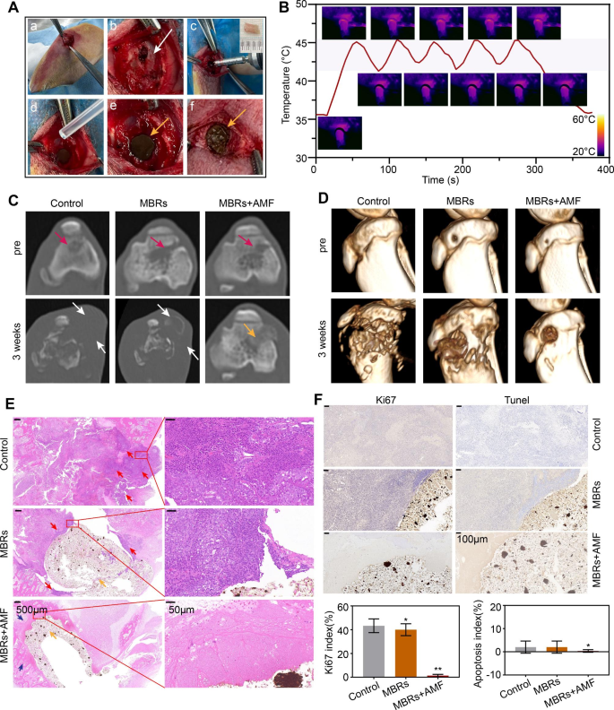
MBRs have proven a very good antitumor impact on 143B human osteosarcoma in a earlier examine, in addition to wonderful osteogenic results in rat cranium defect fashions. Nonetheless, to attain the medical translation of MBRs, it’s nonetheless essential to additional discover the therapy of tumors by simulating the anatomical surroundings of bone tumors. At the moment, in medical observe, surgical resection is the primary alternative for treating tumors that happen in load-bearing bones; nevertheless, surgical procedure can solely take away tumor tissues seen to the bare eye. Due to this fact, multifunctional MBRs not solely exert anti-OS results in a minimally invasive method but in addition help in inhibiting the expansion of residual tumors after surgical therapy, which has necessary significance. We ready a rabbit tibial plateau residual tumor mannequin in situ, as proven in Fig. 7Aa-c. The tumor tissue was partially eliminated with bone. H&E staining confirmed that tumor tissue was distributed round cancellous bone, indicating that solely a part of the tumor tissue was eliminated throughout surgical procedure and that some tumor tissue remained within the tibial plateau (Determine S14). Subsequently, liquid MBRs have been injected into the defect and have become stable by way of liquid trade with surrounding tissues. Surprisingly, MBRs couldn’t solely utterly fill the defect but in addition inhibit bone tissue bleeding by way of liquid trade, decreasing the quantity of bleeding throughout surgical procedure (Fig. 7Advert-f). Then, the tumor website within the rabbit leg was subjected to 5 gentle “on-off” cycles of magnetothermal therapy, and the outcomes indicated that the stuffed MBRs have been evenly heated and had steady magnetothermal properties (Fig. 7B).
Analysis of the MBRs anti-tumor potential for bone tumor in situ. (A) The processes of multinational of a residual tumor mannequin after surgical therapy of tibial plateau bone tumors in rabbits (white arrow: postoperative bone defect, yellow arrows:MBRs). (B) Temperature-time curve of rabbit leg in MBRs group with AMF publicity for 5 on-off cycles and the corresponding infrared thermal pictures. (C) Axial place CT pictures at every follow-up time level of every group (purple arrow: bone tumor in situ, white arrow:bone destruction and swelling of soppy tissue, yellow arrow: injected stable kind MBRs). (D) 3D-reconstructed CT pictures at every follow-up time level. (E) Histological analysis of bone defect and bone tumor after therapy (scale bar is 500 μm and 50 μm, purple arrow: bone tumor in situ, blue arrow: bone tissue after therapy, yellow arrow: injected stable kind MBRs). (F) Immunohistochemical staining (Tunel and Ki67) within the tumor area of every group and corresponding quantitative evaluation (Scale bars: 100 μm). (The information are proven because the means ± SDs, n = 5 per group, n.s. represented no significance and *p < 0.05, **p < 0.01 as compared with the management teams, respectively.)
To judge the expansion of tumors within the tibial plateau, every group of rabbits underwent CT scanning earlier than the intervention and three weeks after therapy. As Fig. 7C reveals, the residual tibial plateau tumors in each the untreated group and the MBR group recurred after 3 weeks, with seen bone destruction and surrounding smooth tissue swelling (white arrows). Nonetheless, within the MBR + AMF group, there was no bone destruction or smooth tissue harm across the tibial plateau. The CT reconstructions (Fig. 7D) confirmed comparable outcomes, with vital bone destruction within the management and MBR teams. H&E staining confirmed a big space of uniformly red-stained cytoplasm, indicating disordered tissue and marked mobile destruction within the MBR + AMF group; the tissues across the MBRs have been homogeneous red-stained supplies with no qualitative construction. Excessive-power microscopy confirmed foam cells, suggesting a response after tumor therapy. Comparatively, many tumor cells have been nonetheless current within the management and MBR teams. Immunohistochemistry (IHC) confirmed that the tumor cells within the management and MBR teams proliferated actively with a low fee of apoptosis, whereas the tumor cells within the MBR + AMF group have been killed and remodeled into an amorphous construction with out nuclei, leading to neither Ki67 nor TUNEL staining. Quantitative correlation evaluation additionally confirmed the identical outcomes, with vital variations within the Ki67 index and apoptosis index between the MBR + AMF group and the management and MBR teams. These outcomes indicated that MBRs can considerably inhibit the expansion of bone tumors in load-bearing bone in situ, additional illustrating the potential of MBRs in mutually-synergistic gentle hyperthermia-starvation remedy for bone tumors in situ.


