IONPs characterization
Ammonia answer was added to the combination of the iron salts (in brilliant yellow) and its shade turned to black, ensuing within the era of iron (III) & iron (II) hydroxides due to the hydrolysis of Fe3+ and Fe2+, respectively. As well as, iron (III) hydroxide was transformed to a different compound (FeOOH) that reacted with Fe(OH)2 to provide Fe3O4. Fe2+ to Fe3+ molar ratio was 1:2 to acquire excessive effectivity in magnetite manufacturing and to forestall the oxidation of Fe2+ to Fe3+. Common reactions within the strategy of Fe3O4 formation are listed beneath (Eqs. (1–4)) [45].
$${Fe}^{3+}left(aqright)+3{OH}^{-}(aq)to Fe(OH{)}_{3}(aq)$$
(1)
$${Feleft(OHright)}_{3}left(aqright)to FeOOHleft(aqright)+{H}_{2}Oleft(lright)$$
(2)
$$F{e}^{2+}left(aqright)+2{OH}^{-}(aq)to {Feleft(OHright)}_{2}(aq)$$
(3)
$$2FeOOHleft(aqright)+Fe(OH{)}_{2}left(aqright)to {Fe}_{3}{O}_{4}left(sright)+2{H}_{2}O(l)$$
(4)
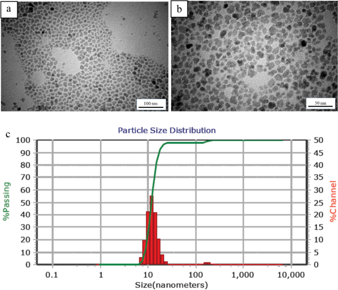
TEM photos of oleic acid-coated magnetite nanoparticles are demonstrated in Fig. 2a, b. These photos point out that near-spherical nanoparticles with uniform measurement (roughly 10 nm) have been synthesized. Additionally it is evident that the floor modification of nanoparticles with oleic acid didn’t meaningfully change the scale of the nanoparticles. DLS evaluation, which is predicated on the Brownian movement of the particles, offers quantitative outcomes of the particle measurement and particle measurement distribution. Determine 2c illustrates a slender measurement distribution within the vary of seven.60–25.55 nm with a mean of 11.8 nm, that’s in good settlement with the TEM outcomes.
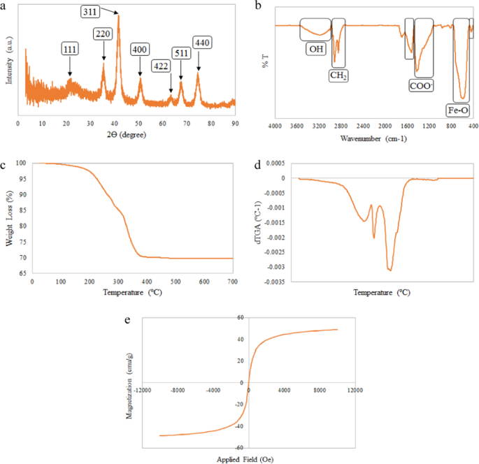
Determine 3a signifies the X-ray diffractogram of IONPs. The XRD sample of the nanoparticles reveals peaks at 2Ɵ = 21.6, 35.3, 41.6, 50.7, 63.3, 67.5, and 74.5° that are attributed to (111), (220), (311), (400), (422), (511), and (440) planes, respectively. The outcomes are in accordance with the magnetite (JCPDS 19–629) reference patterns [46, 47].
Determine 3b exhibits the FTIR spectrum of the modified IONPs. The attribute bands at 444 and 590 cm−1 are assigned to Fe–O bonding [48]. Two attribute bands at 1430 and 1524 cm−1 (as a result of stretching vibration of COO−), affirm the presence of oleic acid on the nanoparticle floor [49]. Different attribute absorption peaks at 2920 and 2850 cm−1 are associated to the uneven and symmetric stretching of CH2 in oleic acid construction, respectively. The bending vibration of the OH band could be attributed to the absorption of water molecules on the floor of the IONPs [46, 49].
TGA evaluation was employed to make sure the floor modification of the IONPs with oleic acid. Determine 3c, d demonstrates the TGA and the corresponding spinoff (dTGA) curves of the coated IONPs. The load loss noticed at temperatures beneath 150 °C was negligible, which is assigned to the evaporation of the adsorbed water. The load loss within the second transition area (temperature vary 210–280 °C) has occurred owing to the elimination of the free or bodily adsorbed oleic acid molecules [50]. The opposite weight reduction noticed at 333 °C is related to the degradation of the oleic acid covalently sure to the IONP floor [51].
Determine 3e illustrates the field-dependent magnetization (M–H) curves of the IONPs. Outcomes revealed the reversible field-dependent magnetization curves with no hysteresis loops, coercivity and remanent magnetization, which demonstrates that the online magnetization of the IONPs is zero within the absence of EMF [52]. These outcomes categorical the super-paramagnetic habits of the synthesized IONPs with excessive saturation magnetization worth of 48.97 emu/g [46].
Ag/IO nanocomposites analysis
FE-SEM picture
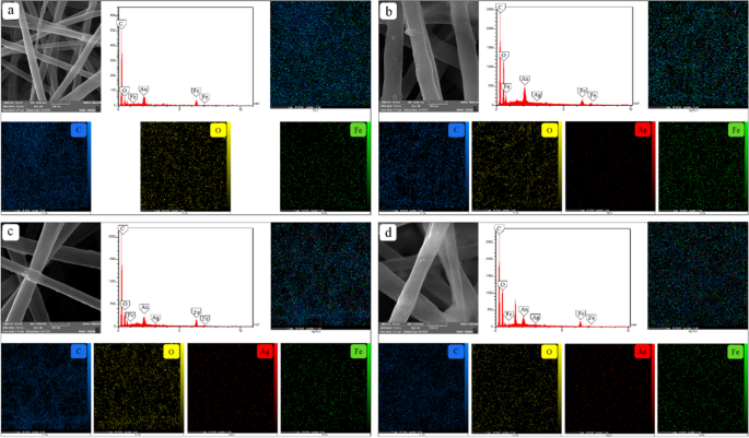
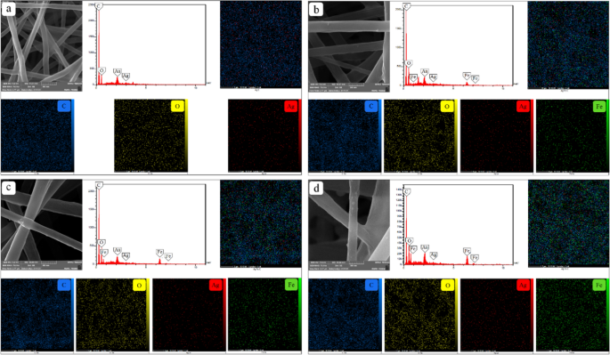
Figures 4 and 5 present the FE-SEM picture, EDX, and the factor maps of Ag and the IONPs within the synthesized nanocomposites. EDX evaluation confirmed the presence of Ag and the IONPs all through the nanocomposites. In keeping with the FE-SEM and the factor map photos, spherical nanoparticles are distributed uniformly among the many fibers with none agglomerations.
Determine 4a–d demonstrates the impact of 4 completely different concentrations of the AgNPs on the nanocomposites containing a hard and fast quantity of the IONPs (10%). In Fig. 4a, C, O, and Fe components could be recognized within the maps. When silver nitrate was added to the composite, Ag additionally appeared within the outcomes. Moreover, as proven within the factor maps of Fig. 4a–d, greater concentrations of silver nitrate within the ready answer results in the elevated content material of AgNPs within the nanocomposite construction. The fixed depth of Fe peaks in EDX evaluation, in addition to the nice distribution of this factor within the maps (a-d), signifies uniform coating of the IONPs in samples 9, 10, 11, and 12. The identical pattern exists in Fig. 5a–d. The small variety of Ag density within the factor maps exhibits the fastened quantity of AgNPs in samples 3, 7, 11, and 15 synthesized utilizing a relentless focus of the silver nitrate within the precursor answer. In proportion to the IONPs focus within the coating answer, the IONPs content material within the composites will increase.
Crystalline construction
Determine 6a demonstrates the X-ray diffractograms of GA/PVA/PCL/Ag and GA/PVA/PCL/Ag/Fe3O4 nanocomposites. The addition of the IONPs to the nanocomposites has decreased the depth of the peaks at 2θ = 24.95 & 27.69°, because of the interplay between PCL and the IONPs [53]. The IONP-containing nanocomposites confirmed three new peaks at 2θ = 34.82, 41.51 and 67.69° (for (220), (311), and (511) planes). Furthermore, the depth of the peaks will increase at 2θ = 50.53 & 74.79° as a result of (400) and (440) planes, respectively.
Chemical construction
FTIR spectra of GA/PVA/PCL/Ag and GA/PVA/PCL/Ag/Fe3O4 nanocomposites are proven in Fig. 6b. The primary distinction is the height at 597 cm−1. This peak is said to the stretching vibration of the steel–oxygen absorption band (Fe–O bond), indicative of the presence of the magnetite nanoparticles. Moreover, interactions between the IONPs and the nanocomposites can change the intensities or the height shift. Within the presence of the IONPs, the peaks of the hydroxyl group, C–H (uneven and stretching), C=O stretching vibration, COO− symmetric stretching, and C–C stretching vibrations have been shifted to 3187, 2927, 1733, 1433, and 846 cm−1, respectively [54].
Magnetic properties
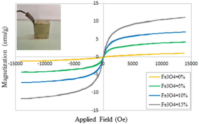
The magnetization–magnetic discipline (M–H) curves of the nanocomposites at 300 Okay are proven in Fig. 7. The zero coercivity and the reversible hysteresis habits of the nanocomposites reveal their super-paramagnetic property, which happens solely on the nanoscale. The saturation magnetization worth of the IONPs was 48.97 emu/g, which was decreased to 4.23, 6.98, and 11.16 emu/g for the nanocomposites containing three completely different concentrations of the IONPs (5, 10, and 15%, respectively). Nevertheless, the outcome additionally reveals super-paramagnetic property [52, 55, 56]. Outcomes exhibit greater saturation magnetization values in comparison with the opposite researchers’ research akin to Ahn and Kang [57]. Utilizing the coating methodology as a substitute of merging the nanoparticles into the answer ready for the electrospinning course of, will increase the particle measurement and improves the magnetic property since magnetic characterization relies on the scale of the synthesized nanoparticles [58].
Antibacterial exercise
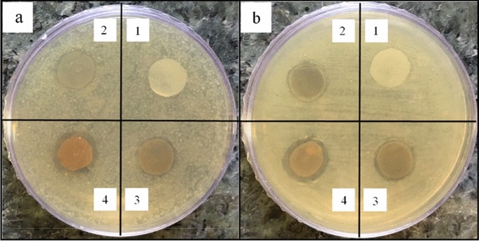
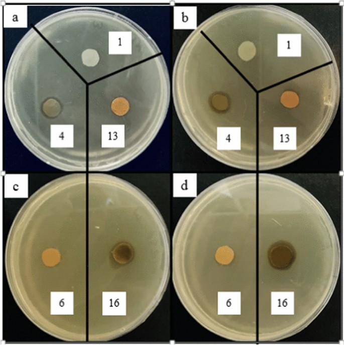
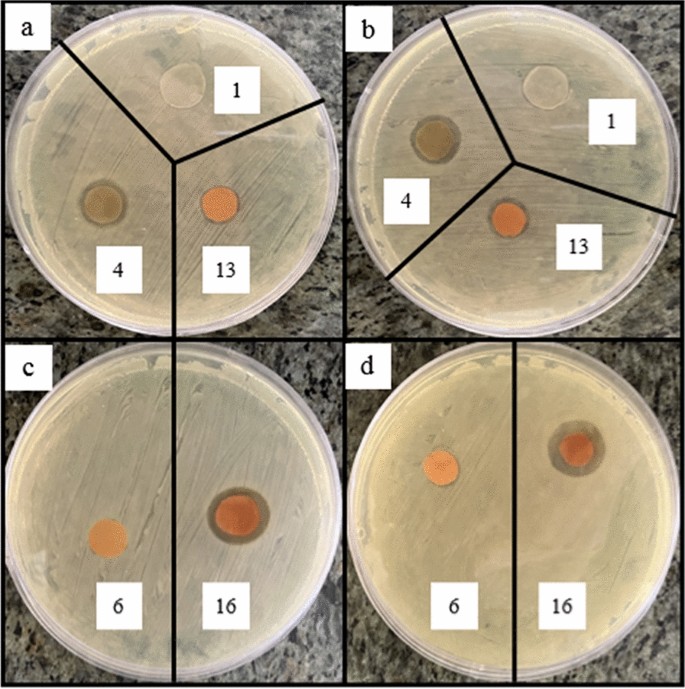
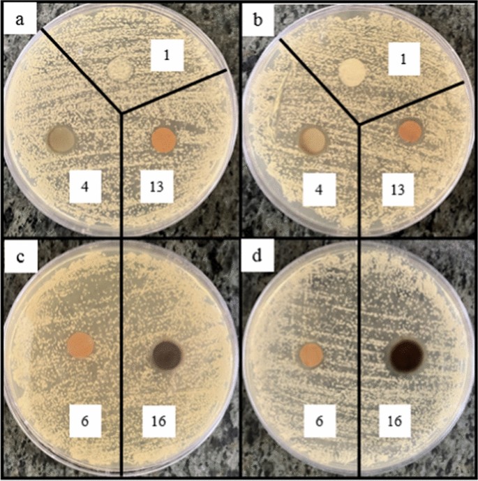
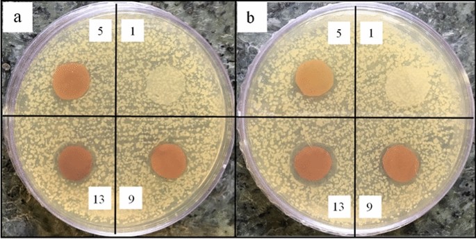
The consequences of AgNPs content material, IONPs focus, and making use of EMF, on the antibacterial exercise of the nanocomposites in opposition to completely different microbial strains (S. aureus and methicillin resistant Staphylococcus aureus (MRSA) (Gram-positive), P. aeruginosa and E. coli (Gram-negative), and C. albicans (Yeast)), have been investigated utilizing two strategies of disk diffusion and colony counting. Resulting from a lot of samples, solely 5 samples embody pattern 1 (management pattern), pattern 4 (containing the very best quantity of silver nanoparticles), pattern 13 (containing the very best quantity of iron oxide nanoparticles), pattern 6 (containing the bottom quantity of each silver nanoparticles and iron oxide) and Pattern 16 (containing the very best quantity of each silver nanoparticles and iron oxide) have been used for microbial strains of MRSA, E. coli, and C. albicans.
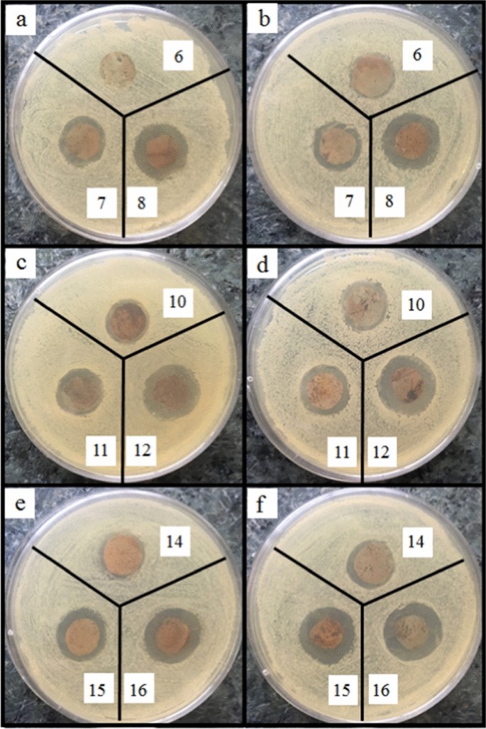
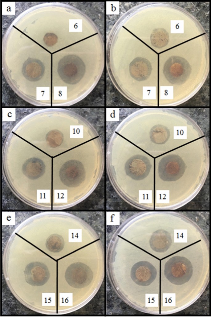
In keeping with Fig. 8a, b, the nanocomposites containing 0.05% AgNPs present low antibacterial exercise. By rising the focus of AgNPs (to 1%), the diameter of the inhibition zone will increase since extra AgNPs could be launched and better antibacterial exercise could be obtained (Figs. 9a, b, 10a, b, 11a, b) [59, 60]. AgNPs adhesion to the cell wall by electrostatic interactions causes its injury. Within the following, it penetrates the cell and pushes the cell content material out, which ends up in DNA deformation and disrupting its transcription and translation, and the era of reactive oxygen species (ROS) and free radicals [13, 61].
The content material of IONPs within the nanocomposite is one other influential issue affecting antibacterial exercise. Figures 9a, b, 10a, b, 11a, b, 12a, b exhibits that rising the IONPs content material from 5 to fifteen% results in elevated antibacterial exercise. The primary mechanism of the antibacterial exercise of IONPs is the oxidative stress attributable to ROS. ROS contain superoxide radicals (O2−), hydroxyl radicals (–OH), singlet oxygen (1O2), and hydrogen peroxide (H2O2), which penetrate the micro organism cells and injury their proteins and DNA [62,63,64]. Then again, micro organism use adhesive floor buildings to connect to tissues. The attachment of IONPs to the cell wall and floor buildings of micro organism causes the bacterial adherence components to be occupied and inactivated, stopping them from binding [65].
A comparability of the photographs in Figs. 9c, d, 10c, d, 11c, d, 13, 14 and the info in Tables 1, 2, 3, 4 and 5 demonstrates that the presence of EMF will increase the antibacterial exercise of IONPs-containing nanocomposites. The magnetic moments of the IONPs are randomly oriented within the absence of EMF. Subsequently, web magnetization is zero. Making use of a powerful sufficient EMF forces the magnetic moments of the IONPs to align alongside the magnetic discipline route [66].
As a result of cheap quantity of the IONPs within the nanocomposites and their superparamagnetic properties, the utilized EMF will increase the IONPs launch and improves the antibacterial exercise. Furthermore, the managed launch of IONPs results in the extra accessible launch of the AgNPs, because of the elevated porosity, and thus the higher antibacterial exercise of the nanocomposites [67, 68].
Along with the disc diffusion assay, the antibacterial exercise of the nanocomposites in opposition to two bacterial strains of S. aureus and P. aeruginosa was additionally carried out by the colony counting methodology, which is proven in Desk 6.
From this desk it’s apparent that the variety of bacterial colonies of petri dishes akin to nanocomposites containing silver and iron oxide nanoparticles for each pathogenic micro organism is considerably decrease than the management nanocomposite (pattern 1). The outcomes present that the addition of silver nanoparticles (pattern 4), the addition of iron oxide nanoparticles (pattern 13), the simultaneous integration of each nanoparticles (samples 6 and 13), and the usage of a magnetic discipline scale back the variety of bacterial colonies. These outcomes are in settlement with agar disk diffusion evaluation.
Among the many microbial strains, P.aeruginosa and MRSA confirmed the very best and lowest sensitivity to the antibacterial nanocomposites, respectively. The noticed variations could be attributed to the presence of a thick layer of peptidoglycan within the cell wall of gram-positive micro organism, which acts like a barrier in opposition to the penetration of the antibacterial nanoparticles into the micro organism and impacts their efficiency [39, 69].
ROS generaion
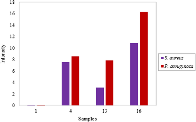
ROS era was measured by the DCFH-DA assay after exposing the nanocomposites to S. aureus and P. aeruginosa to determine whether or not it has an impact on the antibacterial mechanism of Ag/IO nanocomposites or not. The outcomes revealed that NPs-containing nanocomposites, produced extra ROS in comparison with untreated nanocomposite, within the presence of each S. aureus and P. aeruginosa in a concentration-dependent method. Growing the focus of AgNPs to 1% results in about 8% improve in ROS as in comparison with NPs-free nanocomposites. Equally, micro organism uncovered to IONPs-containing nanocomposites demonstrated elevated ROS manufacturing (Fig. 15).
Altogether, the above information recommend that AgNPs act as oxidative stress inducer brokers and trigger the lack of cell viability in S. aureus and P. aeruginosa micro organism by ROS era. ROS formation of AgNPs-containing nanocomposites could be assigned to the upper silver ion launch. The interplay between steel NPs and bacterial cells typically results in ROS manufacturing, which injury to proteins and nucleic acids. The IONPs additionally produce ROS at their floor and rising the focus, causes elevated ROS formation as extra NPs are launched from the nanocomposites [39, 70]. Taken collectively, the above discovering recommend that ROS formation is a attainable mechanism answerable for bacterial cell dying in presence of Ag/IO nanocomposites.
Cytotoxicity
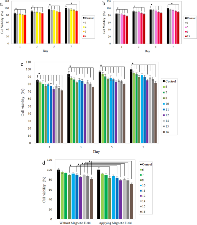
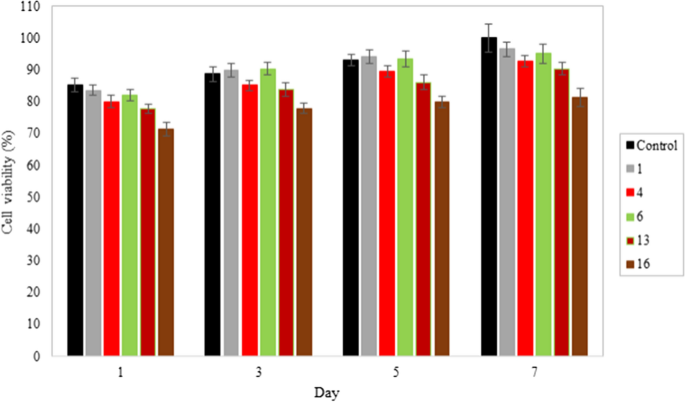
Cytotoxicity assay was carried out to review the biocompatibility of AgNPs, IONPs, and EMF on the fibroblast and macrophage cells. Cells have been cultured on the nanocomposites for 1, 3, 5, and seven days to carry out the MTT assay. Typically, the kind of nanoparticles, their form, measurement, dose, floor properties, and the methods they’re added to the system have an effect on their toxicity [58]. In keeping with Figs. 16a, b and 17, cells have been in a position to develop on all nanocomposites; however rising AgNPs and IONPs contents within the nanocomposites decreased cell proliferation because of the extra launched silver and iron oxide nanoparticles. Outcomes revealed dose-dependent cytotoxicity of the silver nanoparticles [4, 16]. Growing the AgNPs focus to 1% decreased cell viability of fibroblasts and macrophages to about 80%. Ankamwar et al. confirmed that IONPs aren’t poisonous within the focus vary of 0.1–10 µg/mL−1, however cell viability decreases when the focus reaches 100 µg/mL−1 [71] and our research is in settlement with the talked about report. Growing the focus of IONPs from 5 to fifteen% on the primary day, prompted a lower in fibroblast viability from 83.1 to 77.8%. A low focus of IONPs prompted no toxicity in macrophages, even slight development promotion is noticed in prior analysis [72]. On the focus of 15% IONPs phagocytic cells exhibited the next survival price of 85.1% [73]. In keeping with earlier studies, greater concentrations of IONPs may exert cytotoxicity by the era of extreme reactive oxygen species (ROS). Moreover, IONPs impair mobile actions and cell development by adsorbing proteins and different vitamins of the cell tradition medium [72, 74].
MTT assay outcomes of the a IONPs-free nanocomposites containing 0, 0.05, 0.5, and 1% AgNPs; b AgNPs-free nanocomposites containing 0, 5, 10, and 15% IONPs; c Ag/IO nanocomposites containing 0.05, 0.5, and 1% AgNPs; and 5, 10, and 15% IONPs on days 1, 3, 5 and seven; d Ag/IO nanocomposites containing 0.05, 0.5, and 1% AgNPs; and 5, 10, and 15% IONPs on the seventh day within the absence and presence of EMF. Knowledge are reported as imply ± SD (n = 5 and *p < 0.05)
Figures 16c and 17 exhibits the simultaneous impact of AgNPs and IONPs on the biocompatibility of the nanocomposites. As introduced in these determine, when each NPs are built-in into the nanocomposite, their cytotoxicity will increase. Furthermore, the nanocomposites containing excessive concentrations of AgNPs and IONPs on days 1 and three confirmed greater ranges of toxicity, and the cell viability decreased to lower than 80% for each cell varieties. Nevertheless, cell viability was improved even for the nanocomposites containing the utmost quantities of the nanoparticles (pattern 16) due to the appreciable improve within the cell proliferation from day 5. Subsequently, the incorporation of AgNPs and IONPs doesn’t significantly have an effect on the biocompatibility of the nanocomposites.
In keeping with Fig. 16d, making use of an EMF to the nanocomposites with a low focus of the nanoparticles has no appreciable impact on the cell viability, however the nanocomposites containing 15% IONPs launch extra IONPs and AgNPs and consequently improve the system’s cytotoxicity [75].
Typically, because of the bigger measurement of eukaryotic cells than prokaryotes and their structural variations, greater concentrations of nanoparticles are required to trigger cytotoxicity in them. Utilizing nanoparticles in low concentrations which might be efficient in opposition to microorganisms, has no poisonous impact on the eukaryotic cells [76].
Antibiofilm exercise
Greater than 60% of bacterial infections are attributable to the formation of biofilms in continual wounds. In contrast to planktonic micro organism, that are simply killed, biofilms connect to the floor and are proof against a number of antibiotics, which makes their inhibition troublesome. Subsequently, on this research, the inhibitory impact of Ag/IO nanocomposites in opposition to S.aureus and P.aeruginosa biofilms was investigated by a crystal violet staining assay.
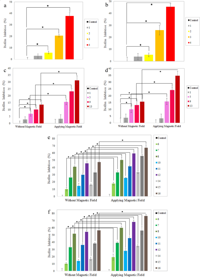
In keeping with Fig. 18a, b, the nanoparticles-free nanocomposites (pattern 1) don’t have any vital impact on the biofilm inhibition, however the addition of small quantities of AgNPs (0.05%) to the nanocomposites, resulted in 6 and 5% biofilm inhibition by S.aureus and P.aeruginosa, respectively; whereas AgNPs content material will increase as much as 1%, promotes the biofilm inhibition to 38 and 45%, respectively [16, 77, 78].
Antibiofilm evaluation of a, b IONPs-free nanocomposites containing 0, 0.05, 0.5, and 1% AgNPs (samples 1, 2, 3, and 4) in opposition to a S. aureus, and b P. aeruginosa; c, d AgNPs-free nanocomposites containing 0, 5, 10, and 15% IONPs (samples 1, 5, 9 and 13) within the absence and presence of EMF in opposition to c S. aureus, and d P. aeruginosa; e, f Ag/IO nanocomposites containing 0.05, 0.5, and 1% AgNPs; and 5, 10, and 15% IONPs within the absence and presence EMF in opposition to e S. aureus, and f P. aeruginosa. Knowledge are reported because the imply ± SD (n = 5 and *p < 0.05)
Determine 18c, d exhibits the impact of various concentrations of IONPs on biofilm inhibition. The nanocomposites containing 5, 10, and 15% IONPs inhibit 7, 10, and 14% of S. aureus biofilm, respectively. The identical pattern was noticed for P. aeruginosa, and with a rise in IONPs content material from 5 to 10% and 15%, biofilm inhibition boosts from 10 to 13% and 15%, respectively [79]. There are a number of mechanisms of biofilm inhibition and antibacterial exercise of IONPs, one in every of which is the era of ROS. As well as, the electrostatic interactions between the nanoparticles and the bacterial cell wall resulting in its destruction and bacterial dying [62].
As introduced in Fig. 18e, f, the biofilm inhibition increments with rising silver and iron oxide nanoparticles. A comparability of the outcomes exhibits that a rise of 0.5% within the quantity of AgNPs has a extra exceptional impact than a rise of 5% within the quantity of IONPs.
A comparability of the nanocomposites manifests that making use of EMF considerably enhances the inhibitory impact because of the elevated launch of IONPs and consequently AgNPs. Because of this, extra AgNPs penetrate the biofilm, resulting in additional inhibition. Along with the antibacterial properties, IONPs within the presence of EMF can mechanically destroy the biofilm construction by penetrating it and displaying extra inhibitory results. Moreover, within the presence of EMF, iron oxide nanoparticles could be extra permeable into the bacterial cell wall by changing the magnetic power into warmth [80]. The best inhibitory exercise of the nanocomposites in opposition to the biofilm of S. aureus and P. aeruginosa was in pattern 16, elevated considerably upon making use of EMF.
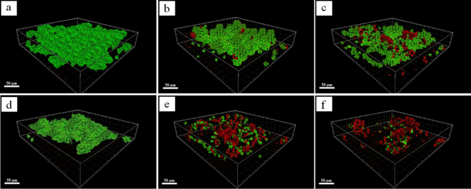
Visible remark of the consequences of the nanocomposite and EMF utility on biofilm inhibition was obtained utilizing CLSM photos (Fig. 19). Samples 6 and 16 have been chosen for biofilm inhibition of S. aurous and P. aeruginosa, respectively. The cells have been stained with SYTO9 (inexperienced) for dwelling cells and propidium iodide (crimson) for lifeless cells.
As proven in Fig. 19a, S. aureus micro organism adhere to the floor, enclosed within the extracellular polymeric substance, and kind a dense and thick biofilm. After therapy of biofilm with pattern 6, a few of the micro organism are killed (Fig. 19b). In actual fact, AgNPs and IONPs, with a excessive surface-to-volume ratio, have been launched, dispersed, and sure to the micro organism, and broken their wall, however the biofilm nonetheless existed and retained its general matrix. By making use of EMF, the diffusivity of the nanoparticles into the biofilm was elevated, which led to a lower within the biofilm density. A comparability of photos (b) and (c) reveals that though the construction of pattern 6 couldn’t kill the micro organism of the biofilm successfully, nanoparticles inhibited the bacterial adhesion and suppressed EPS synthesis [9]. An analogous pattern was additionally noticed for inhibition of P. aeruginosa biofilm. Nevertheless, Ag/IO nanocomposites had a big impact on P. aeruginosa biofilm in comparison with S. aureus. Determine 19d–f demonstrates that after therapy of biofilm with pattern 16, the matrix is broken and just some clusters of bacterial cells are retained. Moreover, when the biofilm is uncovered to EMF, greater than 90% of the biofilm is eradicated and just a few bacterial microcolonies are survived. CLSM outcomes are per the outcomes obtained from the crystal violet staining methodology.
Based mostly on the end result, it may be concluded that though silver is a powerful antibacterial materials, it has low diffusivity in biofilm. In keeping with the scientific studies, utilizing excessive concentrations of silver nanoparticles for additional biofilm inhibition couldn’t be thought of an efficient methodology because of the agglomeration and improve within the measurement of the nanoparticles. This reality is clear within the research carried out by Alharbi et al. (0.5%) [42], Cochis et al. (1.7–1.9%) [81], and Grumezescu et al. (1 mg/cm2) [82], which have used the identical and even greater nanoparticle concentrations than the current research. Then again, the addition of IONPs to AgNPs and utilizing EMF make the silver nanoparticles extra useful and enhance their effectivity for biofilm inhibition.


