Characterization of thermosensitive black phosphorus hydrogels
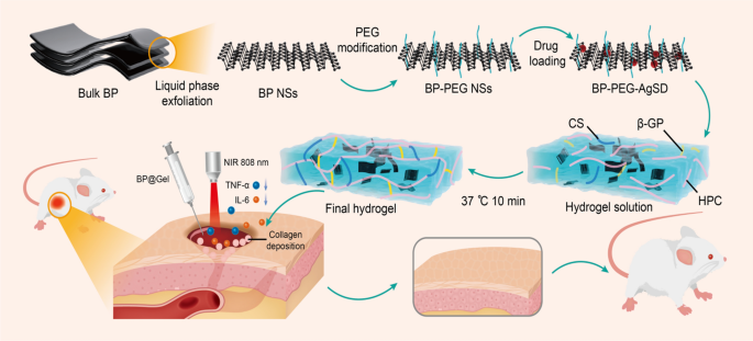
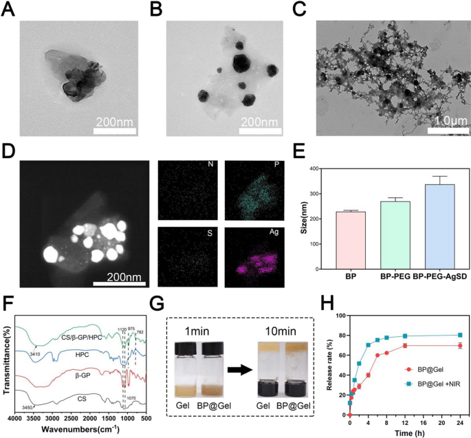
On this examine, black phosphorus nanosheets have been ready by the liquid part exfoliation methodology (Fig. 1) [18]. Transmission electron microscopy, atomic drive microscopy, X-ray photoelectron spectroscopy, and Raman scattering have been used to look at it. The black phosphorus nanosheets have been discovered to have a homogeneous two-dimensional sheet construction, particle dimension of round 220 nm (Fig. 2A), and thickness of roughly 6 nm (Extra file 1: Fig. S3). The XPS peaks of P2p, P2s, C1s, and O1s of black phosphorus nanosheets have been about 130 eV, 200 eV, 290 eV, and 510 eV, respectively (Extra file 1: Fig. S1). Raman scattering revealed three outstanding peaks at 360, 438, and 465 cm (Extra file 1: Fig. S2). The above outcomes confirmed that black phosphorus nanosheets have been efficiently ready. The particle diameter of the black phosphorus nanosheet modified by polyethylene glycol was 266.7 nm and the particle diameter of the black phosphorus nanosheet after drug loading was 370.6 nm. The change within the particle dimension of black phosphorus nanosheets modified by PEG reveals that the modification was comparatively profitable, and the particle dimension change after drug loading confirmed that SSD was loaded on black phosphorus nanosheets (Fig. 2E). Ag and S are the distinctive components of the drug SSD, which could be seen from the mapping diagram of the transmission electron microscope. This additional confirmed that SSD was efficiently loaded (Fig. 2D). Elemental evaluation by transmission electron microscopy confirmed that N, P, S, and Ag accounted for 3.64%, 6.46%, 0.13%, and a couple of.61% of the whole atoms, respectively (Extra file 1: Fig. S4). We additionally investigated the soundness of BP-PEG-AgSD resolution, the colour of BP-PEG-AgSD progressively light on the seventh day, and the absorption spectra of day 0, third day and seventh day confirmed that the absorption depth decreased with time. On the identical time, the storage of BP-PEG-AgSD in PBS elevated its stability, making it an acceptable for the storage of black phosphorus nanosheets (Extra file 1: Fig. S5). Thermosensitive black phosphorus hydrogel (BP@Gel hydrogel) was synthesized utilizing black phosphorus nanosheet loaded with SSD (BP-PEG-AgSD) and chitosan thermosensitive hydrogel (Gel hydrogel), forming a three-dimensional community construction (Fig. 2C).
Characterization of BP@Gel. A TEM picture of BP nanosheets. Scale bar: 200 nm. B TEM picture of BP-PEG-AgSD. Scale bar: 200 nm. C SEM picture of BP@Gel. Scale bar: 1.0 μm. D The scanning transmission map and elemental mapping map of BP-PEG-AgSD. Scale bar: 200 nm. E Particle dimension maps of BP, BP-PEG, and BP-PEG-AgSD nanosheets. F Infrared spectrum of CS/β-GP/HPC hydrogel. G Diagram of the sol–gel part transition of Gel and BP@Gel. H In vitro drug launch price of BP@Gel
To confirm the profitable cross-linking of the hydrogels and that BP-PEG-AgSD had no impact on the chemical composition of the hydrogels, we carried out FT-IR exams on the hydrogels. Infrared spectral options of CS, β-GP, HPC, and CS/β-GP/HPC hydrogels (Fig. 2F). The CS spectrum displayed the polymer’s distinctive absorption peak. The stretching vibration of NH and OH within the CS construction correlated to the absorption peak at 3420 cm−1 [19]. This absorption peak blue-shifted from 3420 to 3410 cm−1 within the CS/β-GP hydrogel spectrum as a result of the amino and hydroxyl teams in CS established hydrogen bonds with the hydroxyl teams in β-GP, reducing the generated by the electron cloud density of NH and OH [20]. Moreover, due to the massive variety of hydroxyl teams in β-GP, the absorption peaks of -OH within the hydrogel at 1120 cm−1 and 1070 cm−1 have been sharper than these of CS. Within the CS/β-GP hydrogel, extra absorption peaks have been current at 975 cm−1 and 782 cm−1, respectively, which represented the symmetry of -GP stretching and bending vibration absorption peaks, along with the absorption peaks present in CS. Based mostly on the modifications in these peaks, we concluded that there have been a whole lot of hydrogen bonds within the CS/β-GP hydrogel. Since CS/β-GP/HPC was a easy bodily combination of HPC, no new absorption peaks appeared within the FTIR spectrum of HPC in contrast with CS/β-GP hydrogel, BP-PEG-AgSD was encapsulated within the hydrogel with out altering the chemical construction (Extra file 1: Fig. S6). After inverting the check tubes, the gel hydrogel and BP@Gel hydrogel teams have been positioned in a relentless temperature incubator at 37 °C. After 1 min, the answer was in a liquid state within the pattern check tubes. Nonetheless, after 10 min, they remodeled right into a gel, which indicated the temperature-sensitive properties of the above hydrogels (Fig. 2G). Usually, the addition of BP-PEG-AgSD didn’t have a serious impression on the gelation time of the hydrogel. Contemplating that the hydrogel could be injected into the pores and skin wound and the physiological temperature is 37 °C, the hydrogel may seal the wound and the loaded medication could possibly be repeatedly launched to advertise wound therapeutic.
The rationale why black phosphorus is loaded with medication is that on the honeycomb construction of black phosphorus nanosheets, after the phosphorus atom varieties a bond with the opposite three phosphorus atoms, there are lone pairs of electrons, that are simply taken away by oxygen molecules, ensuing within the oxidation of black phosphorus nanosheets. Subsequently, the soundness of black phosphorus nanosheets is elevated by blocking its response with oxygen after floor modification with polyethylene glycol. Because of the presence of positively charged phosphate radicals on the floor of black phosphorus nanosheets, they are often mixed with silver sulfadiazine by means of electrostatic adsorption. Chitosan varieties intermolecular hydrogen bonds with sodium glycerophosphate and hydroxypropyl cellulose to type a three-dimensional community construction (Extra file 1: Fig. S7), and black phosphorus nanosheets loaded with silver sulfadiazine are wrapped in hydrogel. The above parts represent the temperature-sensitive black phosphorus hydrogel.
Evaluation of in vitro drug launch
Through the 24 h in vitro drug launch experiment, it was discovered that the in vitro drug launch price of BP@Gel + NIR group was greater than that of the BP@Gel group. The drug launch price of BP@Gel + NIR group was 70.3 ± 1.5% on the 4 h, 75.7 ± 0.6% at 6 h, and 77.7 ± 1.5% at 8 h and tended to be secure; whereas the BP@Gel group reached 80.3 ± 2.1% at 24 h and tended to be secure. The general drug launch price of the BP@Gel + NIR group was greater than that of the BP@Gel group (Fig. 2H).
Adhesion and photothermal evaluation
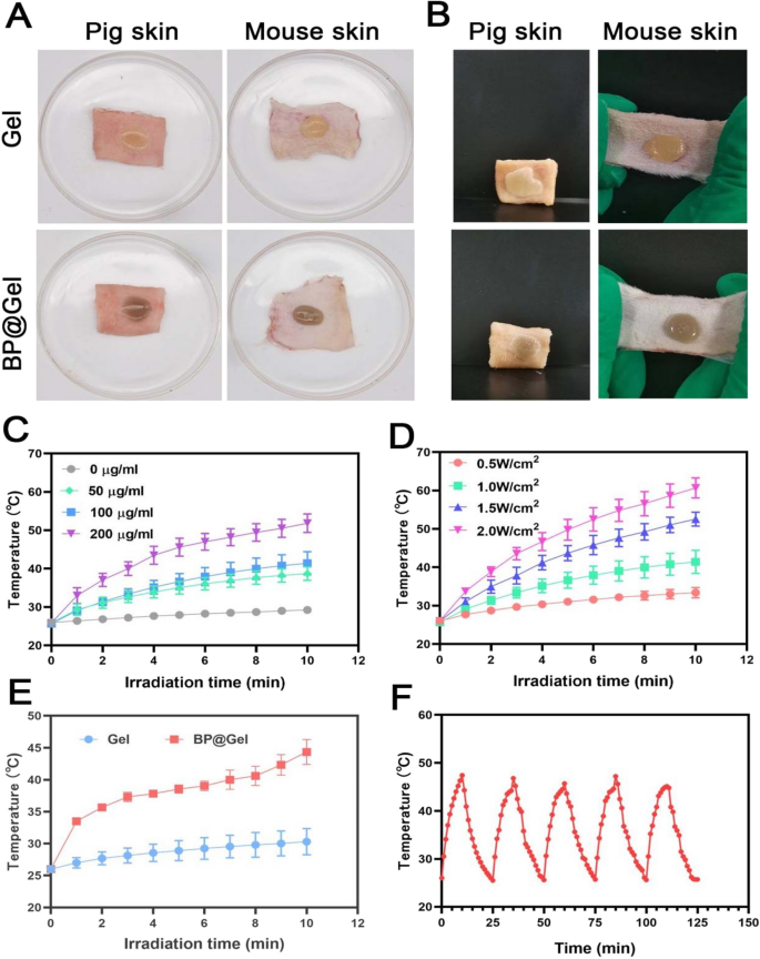
After 10 min, the Gel and BP@Gel shaped a gel, which could possibly be clearly seen within the petri dish, and the Gel and BP@Gel adhered to the mouse pores and skin and pig pores and skin. After stretching, the Gel and BP@Gel nonetheless adhered to the floor of the mouse pores and skin and didn’t fall off. The pig pores and skin was positioned vertically, and the Gel and BP@Gel didn’t fall off, which indicated that BP@Gel had adhered properly to the pores and skin floor (Fig. 3A, B).
With the intention to additional discover the photothermal impact and photothermal stability of black phosphorus nanosheets, the impact of NIR on totally different concentrations of black phosphorus nanosheets was evaluated on this experiment. Because the focus of black phosphorus nanosheets elevated, the temperature elevated, and the dispersion of 200 μg/mL black phosphorus nanosheets lastly reached 63.6 °C at 1 W/cm2 (Fig. 3C). The focus of the Gel group was 100 μg/mL Beneath totally different energy irradiation, as the facility elevated, the temperature additionally elevated (Fig. 3D). The Gel group and BP@Gel group have been irradiated with NIR with an influence density of 1.0 W/cm2 for 10 min, and the temperature modifications have been recorded by a thermocouple thermometer. After 10 min of irradiation, the ultimate temperature of BP@Gel reached 45 °C. In distinction, nearly no vital temperature change was noticed within the Gel group, which indicated that the Gel itself had no photothermal impact. When BP-PEG-AgSD resolution was added and irradiated with NIR with an influence density of 1.0 W/cm2, the temperature elevated with longer irradiation time (Fig. 3E).
Photothermal stability is without doubt one of the vital elements to guage the impact of photothermal remedy. Poor photothermal stability limits the appliance of many biomaterials with photothermal properties within the biomedical discipline. With the intention to consider the photothermal stability of BP@Gel, 5 consecutive NIR mild cycles have been carried out utilizing the switching mode. The temperature of BP@Gel elevated quickly inside 10 min, and after turning off the laser for 15 min, the BP@Gel cooled down and reached the preliminary temperature. In 5 consecutive NIR (irradiation-cooling) cycles, it was discovered that the utmost photothermal conversion temperature of BP@Gel didn’t change considerably, which instructed that BP@Gel had good photothermal stability. Based mostly on the superb photothermal impact and photothermal stability of BP@Gel, BP@Gel could function a promising candidate materials for PTT (Fig. 3F).
Adhesion and photothermal properties of BP@Gel. A Hydrogel unfold on pig pores and skin and mouse pores and skin. B Vertical pig pores and skin and stretched mouse pores and skin.C Temperature variation curves of black phosphorus nanosheet dispersions with totally different concentrations underneath near-infrared (1W/cm2) irradiation. D 100 μg/mL black phosphorus nanosheet dispersion Temperature change of liquid underneath near-infrared irradiation with totally different powers. E Temperature change of hydrogel underneath near-infrared (1W/cm2) irradiation. F 5 cycles of heating and cooling of BP@Gel
Analysis of hemocompatibility and cytocompatibility in vitro
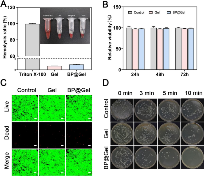
Nanocomposite hydrogels have to be biocompatible earlier than they can be utilized within the clinic. Hemolysis assay is used to evaluate the cytotoxicity of medicinal brokers and can also be used to characterize blood contact supplies. Based on the American Society for Testing and Materials Designation (ASTMF-756-00), a hemolysis price (HR) better than 5% is taken into account as extreme hemolysis, whereas a hemolysis price lower than 2% and between 2 and 5% is deemed non-hemolysis, and gentle hemolysis, respectively [21]. The hemocompatibility of BP@Gel hydrogel was quantified utilizing a hemolysis check, with Triton X-100 as a constructive management and PBS as a destructive management [22]. The constructive management group’s supernatant was vivid purple, with no sediment on the backside, indicating that the cells have been destroyed and extreme hemolysis had occurred, whereas the destructive management group, Gel group, and BP@Gel hydrogel group’s supernatants have been clear and clear, with a big pellet of purple blood cells on the backside. The HR of the hydrogels within the Gel hydrogel group and the BP@Gel hydrogel group have been each lower than 3% (Fig. 4A), exhibiting that the hydrogels have been hemocompatible.
NIH3T3 cells and hydrogel extracts have been used as in vitro fashions for the viability and stay/lifeless fluorescence labeling experiments. First, NIH3T3 cells have been co-cultured with varied doses of hydrogel extracts for twenty-four h, 48 h, and 72 h, and the OD worth was measured utilizing the CCK8 assay. The cell viability of every group was greater than 85% at 24 h, 48 h, and 72 h, and the cell viability at totally different concentrations of hydrogel extracts was better than 85% in any respect the examined time factors (Fig. 4B, Extra file 1: Fig. S8). For the stay/lifeless cell staining, the cells have been co-cultured with the hydrogel extract for 72 h earlier than being labeled with the stay/lifeless reagents and visualized utilizing a confocal microscope (Leica TCS SP8) (Fig. 4C). The outcomes revealed that the Gel hydrogel group had a small variety of lifeless cells (purple), with most of them being stay cells (inexperienced). The morphology of stay cells within the BP@Gel hydrogel group was modified from irregular polygons or triangles to circles after 72 h because of the addition of black phosphorus nanosheets, whereas the variety of lifeless cells elevated. Usually, the viability of NIH-3T3 cells in every experimental group was excessive, indicating good cytocompatibility of the hydrogel.
Antimicrobial properties of the hydrogels
On this examine, a novel BP@Gel hydrogel-based wound dressing with antibacterial exercise was ready. Utilizing the plate unfold methodology, Staphylococcus aureus (Gram-positive micro organism) was chosen as a consultant mannequin of micro organism. The bacterial viability of Staphylococcus aureus was considerably decreased after incubation with BP@Gel hydrogel underneath NIR mild irradiation. The Gel hydrogel group alone didn’t present this antibacterial impact on Staphylococcus aureus. Based mostly on the outcomes of the Management group, when NIR mild was irradiated for 10 min, it may kill some micro organism. On the identical time, the Gel group had an antibacterial impact at 10 min. It could possibly be that chitosan itself possessed antibacterial properties and will inhibit the expansion of micro organism. When NIR radiation was utilized for 10 min, the BP@Gel group had a major antibacterial impact in comparison with the Management group and the Gel group. On the identical time, the antibacterial impact of the BP@Gel group decreased with out NIR irradiation. The above outcomes confirmed that the mixed utility of NIR irradiation and hydrogel may obtain synergistic antibacterial results and that the mixture of BP@Gel and NIR irradiation was sufficient to kill a lot of the micro organism (Fig. 4D).
Analysis of wound therapeutic in in vivo mouse fashions of pores and skin wound
Hydrogels are generally used as pores and skin wound dressings to maintain wounds moist and promote tissue therapeutic [23, 24]. Wound therapeutic is a dynamic, well-controlled course of that entails partially overlapping levels, together with hemostasis, irritation, proliferation, and reworking [25, 26]. The antibacterial agent loaded onto the black phosphorus nanosheets on this examine was SSD. SSD is an FDA-approved topical antibiotic that has been reported to be efficient in opposition to Gram-negative and Gram-positive micro organism [27]. As well as, SSD possesses wound-healing impact [28,29,30].
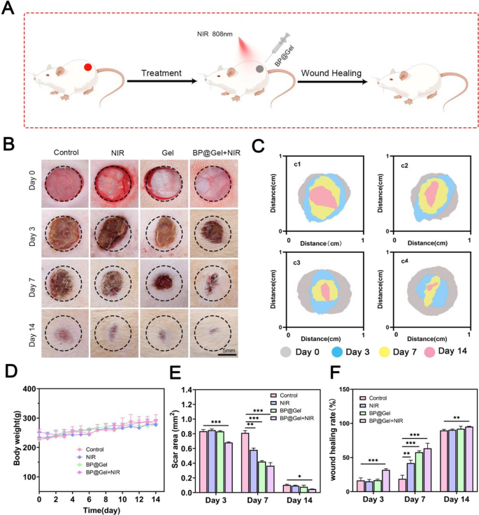
The rat pores and skin full thickness wound mannequin was used to guage the in vivo wound therapeutic properties of thermosensitive black phosphorus hydrogel as a possible wound dressing. A round full-thickness pores and skin defect wound with a diameter of 1 cm was generated on the again of SD rats. BP@Gel hydrogel was then injected into the wound and irradiated with 808 nm near-infrared mild for 10 min to advertise wound therapeutic (Fig. 5A). After treating the injuries in numerous teams, wound therapeutic was evaluated (Fig. 5B, C). On the third day, the wound space of the BP@Gel hydrogel group was discovered to be considerably smaller than that of the opposite teams. On day 7, the management group had some exudate together with redness and swelling, whereas the wound within the BP@Gel hydrogel group had nearly no swelling or redness. This indicated that BP@Gel hydrogel was in a position to cut back wound irritation, which was doubtlessly mediated by the sustained launch of black phosphorus nanosheets and SSD from the hydrogels. The separate Gel group containing chitosan possessed wound therapeutic skill, enhanced wound pores and skin metabolism, lined and remoted ulcer wounds, inhibited bacterial development and guarded wounds, and inhibited scar formation [31, 32]. We confirmed that the BP@Gel hydrogel launched black phosphorus nanosheets and SSD concurrently, which was more practical than the Gel hydrogel group alone, and the Gel hydrogel group containing chitosan additionally promoted wound therapeutic. On day 14, the injuries within the totally different teams have been principally healed, however these within the BP@Gel hydrogel group healed higher (Fig. 5E, F). From day 0 to day 14, the physique weight of the rats in numerous teams progressively elevated, indicating that the therapies in numerous teams didn’t change the physique weight of the rats (Fig. 5D). General, the BP@Gel hydrogel group had the smallest scar space and the perfect pores and skin regeneration skill in contrast with the opposite experimental teams.
Results of thermosensitive black phosphorus hydrogel on wound therapeutic in vivo. A Schematic illustration of hydrogel selling wound therapeutic. B Wound photographs of various teams after the wound space was handled on days 0, 3, 7, and 14 after surgical resection. Scale bar: 5 mm. C Schematic diagram of wound therapeutic simulated by picture software program in Management group (c1), NIR group (c2), BP@Gel group (c3) and BP@Gel + NIR group (c4). D Graph of physique weight change in rats throughout wound therapeutic. E Wound space map. F Graph of wound therapeutic price. (*P < 0.05, **P < 0.01, ***P < 0.001)
Histological evaluation of the regenerated tissue
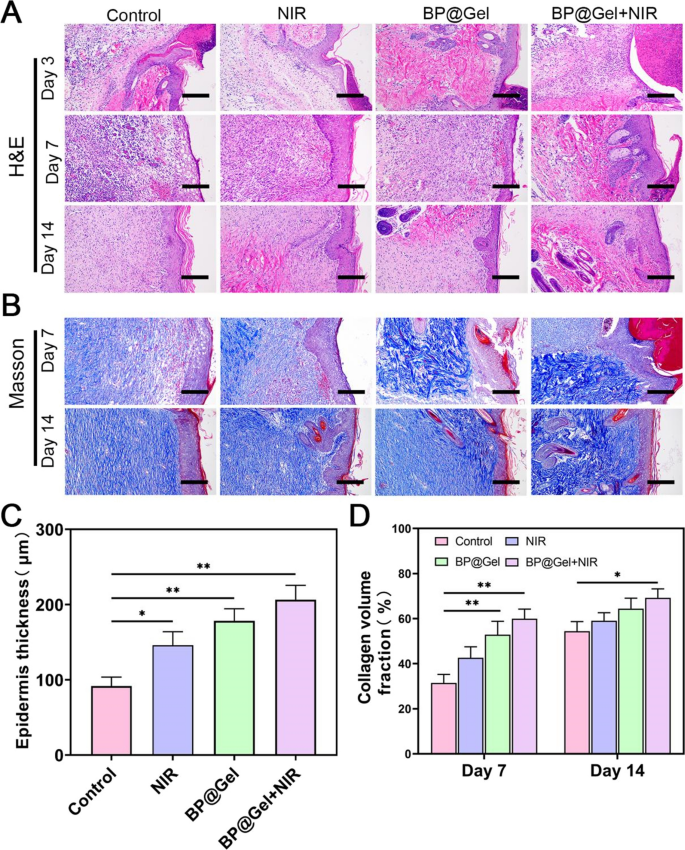
Histological analysis elucidates the tissue regeneration course of and divulges the therapeutic impact of the hydrogel on pores and skin wounds [33, 34]. To evaluate wound therapeutic, HE staining was performed on days 3, 7, and 14. On day 7, the dermis of the NIR group consisted of a considerable variety of inflammatory cells penetrating the granulation tissue, however the wounds within the Gel hydrogel group and the BP@Gel hydrogel group contained fewer inflammatory cells and established an epithelial layer. This was doubtlessly because of the presence of chitosan within the Gel hydrogel group, which promoted wound therapeutic, and on the identical time, SSD loaded on the black phosphorus nanosheets promoted antibacterial results and synergized with photothermal results from the nanosheets. It was beforehand proven that black phosphorus nanosheets coated with silver nanoparticles have been extraordinarily environment friendly at inducing antibacterial results in vivo [35]. The pores and skin tissue construction of the BP@Gel hydrogel group was regular on the 14th day, as have been the morphology and construction of the pores and skin appendages, and there was no inflammatory cell infiltration within the tissue (Fig. 6A). Moreover, the thickness of the newly created dermis elevated to various levels in every group on day 7, with the thickness of the Management group, NIR group, Gel group, and BP@Gel + NIR group being 91.76, 146.03, 178.27, and 206.47 μm, respectively (Fig. 6C). The elemental construction of the dermis and dermis have been current on the wound floor of the Management group on day 14, however the dermis was nonetheless within the means of restore, the inflammatory response was gentle and no pores and skin appendages have been detected. The pores and skin construction was barely aberrant within the NIR group, and a few collagen fibers within the dermis have been absent. In distinction, the connective tissue and epithelial cells within the Gel hydrogel and BP@Gel hydrogel teams appeared regular with extra fibroblasts, new blood vessels, hair follicles, the collagen fibers within the dermis have been neatly organized, and the morphology and construction of the pores and skin appendages have been additionally regular. There was no main inflammatory response within the two teams, indicating that the 2 hydrogels possessed good wound therapeutic properties. On day 14, the BP@Gel hydrogel group confirmed the perfect wound restore end result, with extra fibroblasts and hair follicles than within the different three teams.
Histological evaluation of pores and skin wounds in numerous teams on day 3, day 7 and day 14. A HE staining and B Masson staining outcomes of pores and skin tissue. Scale bar: 100 μm. C Epidermal thickness map at day 7. D Masson staining quantitative evaluation of collagen fiber content material (%) in numerous teams. (*P < 0.05, **P < 0.01)
Masson staining was carried out to guage collagen fibers throughout wound therapeutic [36]. On days 7 and 14, all teams confirmed collagen deposition. The management group confirmed much less collagen deposition on the wound floor than the opposite teams. The collagen fibers have been denser, thicker and higher ordered within the BP@Gel hydrogel group, which promoted wound therapeutic with the best collagen density and probably the most orderly fiber construction, akin to pure pores and skin (Fig. 6B). The collagen quantity % was biggest within the BP@Gel hydrogel group (Fig. 6D). The above outcomes demonstrated that BP@Gel hydrogel enormously elevated collagen deposition throughout wound therapeutic.
IL-6, TNF-α, CD31, and CD68 expression throughout wound therapeutic
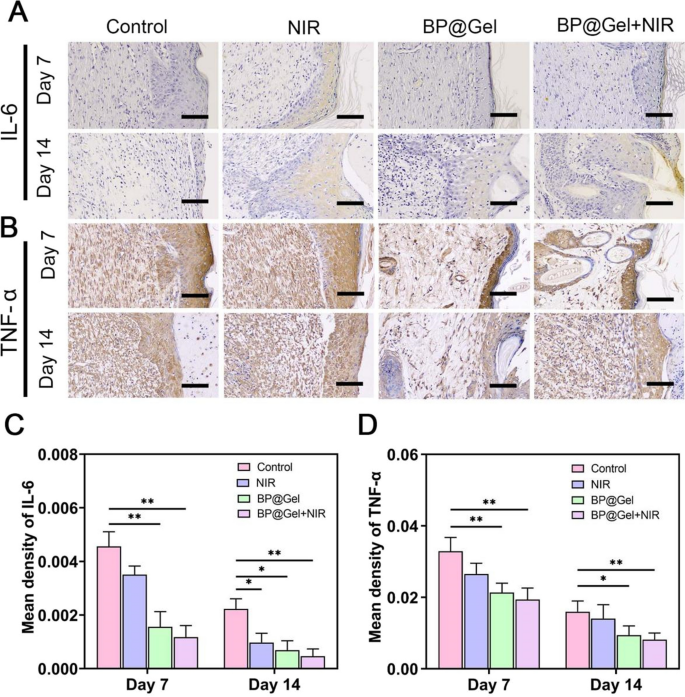
To additional examine the mechanism of BP@Gel hydrogel mediated wound therapeutic, the elements influencing the wound therapeutic course of have been examined, notably the expression of inflammatory angiogenic elements. IL-6 and TNF-α are typical pro-inflammatory cytokines concerned within the early stage of wound therapeutic when the pores and skin wound is within the inflammatory part, and they’re important markers of inflammatory response [37,38,39,40]. There was a really low expression of IL-6 and TNF-α within the BP@Gel hydrogel group in contrast with the opposite teams (Fig. 7A–D), which was much like the outcomes from HE staining (Fig. 6A). This indicated that BP@Gel hydrogel had good anti-inflammatory properties.
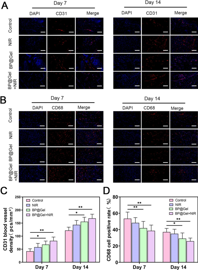
As well as, the expression of cytokines related to irritation (CD68) and angiogenesis (CD31) have been evaluated throughout pores and skin regeneration [41, 42]. Wound neovascularization was examined by immunofluorescent labeling with CD31 after 7 and 14 days of remedy. The injuries handled by the Gel group and the BP@Gel hydrogel group confirmed greater CD31 expression, and the CD31 blood vessel density elevated dramatically (Fig. 8C), demonstrating that the BP@Gel hydrogel promoted wound closure and induced angiogenesis. Equally, immunofluorescence staining for CD68 was decrease within the BP@Gel hydrogel group in comparison with the opposite teams (Fig. 8D). Usually, remedy with BP@Gel hydrogel induced angiogenesis and inhibited inflammatory cytokine expression throughout the wound therapeutic course of.
In vivo biocompatibility of thermosensitive black phosphorus hydrogel
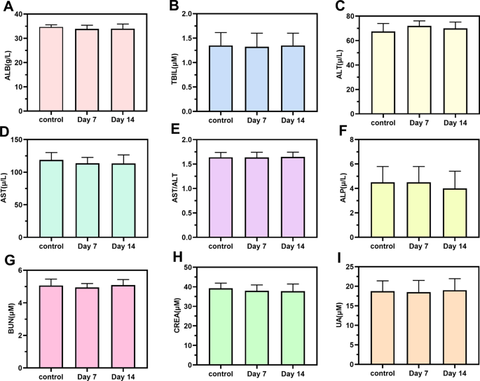
Biosafety is a vital criterion for the appliance of nanomaterials in biomedicine. The in vivo biocompatibility of temperature-sensitive black phosphorus hydrogel was comprehensively analyzed to make sure their in vivo security. On days 7 and 14 following dosing, blood biochemical markers have been examined. As proven in Fig. 9A–I, blood biochemical evaluation revealed that albumin (ALB), alanine transferase (ALT), aspartate transferase (AST), aspartate/glucoside (AST/ALT), alkaline phosphatase (ALP), blood urea nitrogen (BUN), creatinine (CREA), uric acid (UN), and different main blood parameters weren’t considerably totally different between the management group and the experimental group. Concurrently, HE staining was performed for histological analysis of the first organs of rats, together with the center, liver, kidney, spleen, lung and mind, with a view to additional analyze the toxicity of BP@Gel to near-infrared-irradiated rats. Histological examination revealed no variations between the totally different teams, indicating the in vivo security of BP@Gel (Extra file 1: Fig. S9). The BP@Gel discharged on the wound web site didn’t promote systemic toxicity, which indicated that the thermosensitive black phosphorus hydrogel had excessive biocompatibility in vivo and was appropriate as a brand new wound dressing to advertise wound therapeutic.


