| Sep 14, 2023 |
|
|
|
(Nanowerk Information) The truth that people and different dwelling organisms can develop and develop from a single cell depends on a course of referred to as embryonic improvement. For wholesome tissue to type, cells within the embryo have to prepare themselves in the proper method in the proper place on the proper time. When this course of doesn’t go proper, it can lead to beginning defects, impaired tissue regeneration or most cancers. All of which makes understanding how totally different cell sorts manage into a fancy tissue structure one of the vital basic questions in developmental biology.
|
|
Whereas researchers are nonetheless far from absolutely understanding the method, a gaggle of Brown College scientists has spent the previous handful of years serving to the sector inch nearer. Their secret? A department of arithmetic referred to as topology.
|
|
The analysis staff at Brown, made up of biomedical engineers and utilized mathematicians, created a machine studying algorithm utilizing computational topology that profiles shapes and spatial patterns in embryos to review how these cells manage themselves into tissue-like architectures. In a brand new research, they take that system to the following degree, opening a path to learning how a number of forms of cells assemble themselves.
|
|
The work is described in npj Techniques Biology and Functions (“Topological information evaluation of spatial patterning in heterogeneous cell populations: clustering and sorting with various cell-cell adhesion”).
|
|
“In tissues, there could also be variations in how one cell adheres to the identical cell sort, relative to the way it adheres to a distinct cell sort,” mentioned Ian Y. Wong, an affiliate professor in Brown’s College of Engineering who helped develop the algorithm. “There’s this attention-grabbing query of how these cells know precisely the place to finish up inside a given tissue, which is commonly spatially compartmentalized into distinct areas.”
|
|
For instance, in an animal embryo, the outer layer of cells goes on to type pores and skin, the center layer kinds muscle and bone, whereas the innermost layer kinds the liver or lungs. Cells inside every layer will preferentially adhere to one another, sorting aside from cells in different layers that go on to type different components of the physique.
|
|
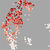
Within the Seventies, scientists found that cells inside frog embryos could possibly be gently separated aside and once they had been blended again collectively, they’d spontaneously rearrange into their preliminary group. This happens as a result of the cells have totally different affinities for one another, and as they assemble and cluster, sure topological patterns of linkages and loops are preserved.
|
|
“Within the context of those spatial preparations of tissues, you possibly can study loads from what’s there, but in addition from what’s not there on the similar time,” mentioned Dhananjay Bhaskar, a current Brown Ph.D. graduate who led the work and is now a postdoctoral researcher at Yale College.
|
|
The Brown researchers confirmed in 2021 (Gentle Matter, “Topological information evaluation of collective and particular person epithelial cells utilizing persistent homology of loops”) how their strategy can profile the topological traits of 1 cell sort that organizes into totally different spatial configurations and will make predictions on it.
|
|
The hiccup with the unique system was that it’s a sluggish and labor-intensive course of. The algorithm painstakingly in contrast these topological options one-by-one towards these in different units of cell positions to find out how topologically totally different or related they’re. The method took a number of hours and, primarily, held the algorithm again from its full potential in understanding how cells assemble themselves, and from with the ability to simply and precisely examine what occurs when situations change — a key in breaking down what occurs when issues go mistaken.
|
|
Within the new research, the analysis staff begins to deal with that limitation with what’s referred to as persistence photos. These photos are a standardized picture-like format for representing topological options, enabling fast comparability throughout giant datasets of cell positions.
|
|
They then used these photos to coach different algorithms to generate “digital fingerprints” that seize the important thing topological options of the info. This drives down the computation time from hours to seconds, enabling the researchers to check hundreds of simulations of cell group through the use of the fingerprints to categorise them into related patterns with out human enter.
|
|
The researchers say the purpose is to work backward and infer the foundations that describe how totally different cell sorts prepare themselves based mostly on the ultimate sample. As an example, in the event that they tinker with how sure cells are extra adhesive or much less adhesive, the researchers can establish how and when dramatic alterations happen in tissue structure.
|
|
The strategy has the potential to be utilized to understanding what occurs when the developmental course of goes off monitor and for laboratory experiments testing how totally different medicine can alter cell migration and adhesion.
|
|
“In case you can see a sure sample, we are able to use our algorithm to inform why that sample emerges,” Bhaskar mentioned. “In a method, it’s telling us the foundations of the sport in the case of cells assembling themselves.”
|
